

perturbation in endogenous pathways by the biosensor itself while probing target;
thus, they allow fluorescence imaging with a closer representation to the in vivo
system over a longer period of time [50]. On the other hand, fluorescent dyes are
limited by their stability, photobleaching, and cytotoxicity, which are often driven by
their non-native interaction with biomolecules that interferes with endogenous fate
of the analytes, and thus might limit relevance of such a study to single time point
analysis alone [51]. Furthermore, unlike dyes, GES are not prone to leakage during
long-term experiments and offer high-throughput screening in drug development.
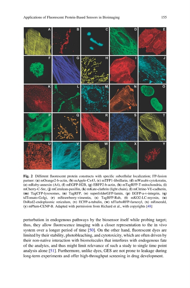
Fig. 2 Different fluorescent protein constructs with specific subcellular localization; FP-fusion
partner: (a) mOrange2-b-actin, (b) mApple-Cx43, (c) mTFP1-fibrillarin, (d) mWasabi-cytokeratin,
(e) mRuby-annexin (A4), (f) mEGFP-H2B, (g) EBFP2-b-actin, (h) mTagRFP-T-mitochondria, (i)
mCherry-C-Src, (j) mCerulean-paxillin, (k) mKate-clathrin (light chain), (l) mCitrine-VE-cadherin,
(m) TagCFP-lysosomes, (n) TagRFP, (o) superfolderGFP-lamin, (p) EGFP-a-v-integrin, (q)
tdTomato-Golgi, (r) mStrawberry-vimentin, (s) TagBFP-Rab, (t) mKO2-LC-myosin, (u)
DsRed2-endoplasmic reticulum, (v) ECFP-a-tubulin, (w) tdTurboRFP-farnesyl, (x) mEmerald,
(y) mPlum-CENP-B. Adapted with permission from Richard et al., with copyrights [48]
Applications of Fluorescent Protein-Based Sensors in Bioimaging
155
thus, they allow fluorescence imaging with a closer representation to the in vivo
system over a longer period of time [50]. On the other hand, fluorescent dyes are
limited by their stability, photobleaching, and cytotoxicity, which are often driven by
their non-native interaction with biomolecules that interferes with endogenous fate
of the analytes, and thus might limit relevance of such a study to single time point
analysis alone [51]. Furthermore, unlike dyes, GES are not prone to leakage during
long-term experiments and offer high-throughput screening in drug development.
Fig. 2 Different fluorescent protein constructs with specific subcellular localization; FP-fusion
partner: (a) mOrange2-b-actin, (b) mApple-Cx43, (c) mTFP1-fibrillarin, (d) mWasabi-cytokeratin,
(e) mRuby-annexin (A4), (f) mEGFP-H2B, (g) EBFP2-b-actin, (h) mTagRFP-T-mitochondria, (i)
mCherry-C-Src, (j) mCerulean-paxillin, (k) mKate-clathrin (light chain), (l) mCitrine-VE-cadherin,
(m) TagCFP-lysosomes, (n) TagRFP, (o) superfolderGFP-lamin, (p) EGFP-a-v-integrin, (q)
tdTomato-Golgi, (r) mStrawberry-vimentin, (s) TagBFP-Rab, (t) mKO2-LC-myosin, (u)
DsRed2-endoplasmic reticulum, (v) ECFP-a-tubulin, (w) tdTurboRFP-farnesyl, (x) mEmerald,
(y) mPlum-CENP-B. Adapted with permission from Richard et al., with copyrights [48]
Applications of Fluorescent Protein-Based Sensors in Bioimaging
155