

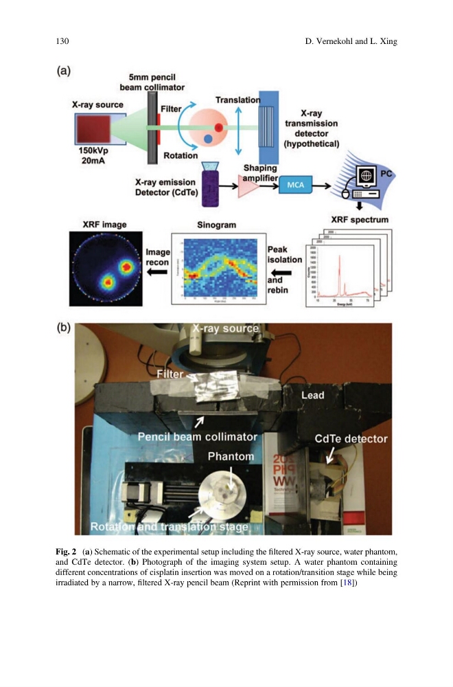
Fig. 2 (a) Schematic of the experimental setup including the filtered X-ray source, water phantom,
and CdTe detector. (b) Photograph of the imaging system setup. A water phantom containing
different concentrations of cisplatin insertion was moved on a rotation/transition stage while being
irradiated by a narrow, filtered X-ray pencil beam (Reprint with permission from [18])
130
D. Vernekohl and L. Xing
and CdTe detector. (b) Photograph of the imaging system setup. A water phantom containing
different concentrations of cisplatin insertion was moved on a rotation/transition stage while being
irradiated by a narrow, filtered X-ray pencil beam (Reprint with permission from [18])
130
D. Vernekohl and L. Xing