

The contrast in XLCT imaging relies on the excitation of nanophosphors. These
nanophosphors usually consist of a nanoparticle made from a high atomic number
element, and the particle is bounded to phosphor groups which cause the emission of
optical or near-infrared (NIR) photons. The emission of these photons is stimulated
by a high-energy electron ejected during a Compton or photoelectric interaction of
the exciting X-ray with the high atomic number element. The optical signal can be
measured with cost-efficient CCD cameras, while NIR signals require more sophisticated detection hardware. The contrast agents used in XLCT are partly translated
from tracers used in laser stimulated NIR optical imaging. These probes which emit
light with a higher wavelength than optical photons can penetrate deeper into soft
tissue. XLCT imaging promises to surpass the depth-dependent restrictions of
optical imaging even further as the X-rays can even excite contrast agents in very
thick samples. Furthermore, X-rays produce no autofluorescent signal in normal
tissue which is a problem in laser-excited optical imaging. These advantages will
extend the applicability of tracers built from rare-earth nanophosphors in medical
imaging.
Both XFCT and XLCT do not require X-ray detectors in the transmission
direction. Hence, traditional CT imaging systems can be extended by XF or XL
detection systems, providing X-ray CT with the ability to see molecular contrasts
[4, 6, 8–10]. The information undetectable by X-ray attenuation (such as cellular and
molecular activities and physiologic function) is critical to detect a disease at its early
malignant stage, to differentiate between aggressive and non-aggressive phenotypes,
and to understand how to treat a disease effectively [11–13].
The extension of XF imaging to XFCT is, to a large extent, analogous to the
development of magnetic resonance imaging (MRI) from nuclear magnetic resonance (NMR) in the 1970s, which has fundamentally changed medical practice.
Indeed, if the detection limit of XF analysis (1–10 ppm) can be fully realized in
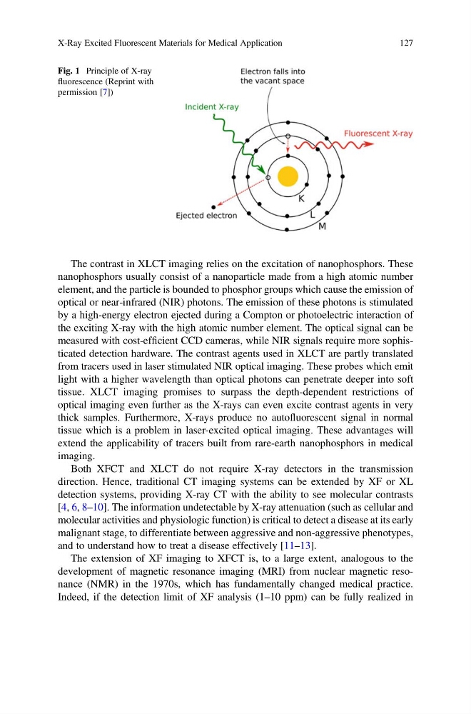
Fig. 1 Principle of X-ray
fluorescence (Reprint with
permission [7])
X-Ray Excited Fluorescent Materials for Medical Application
127
nanophosphors usually consist of a nanoparticle made from a high atomic number
element, and the particle is bounded to phosphor groups which cause the emission of
optical or near-infrared (NIR) photons. The emission of these photons is stimulated
by a high-energy electron ejected during a Compton or photoelectric interaction of
the exciting X-ray with the high atomic number element. The optical signal can be
measured with cost-efficient CCD cameras, while NIR signals require more sophisticated detection hardware. The contrast agents used in XLCT are partly translated
from tracers used in laser stimulated NIR optical imaging. These probes which emit
light with a higher wavelength than optical photons can penetrate deeper into soft
tissue. XLCT imaging promises to surpass the depth-dependent restrictions of
optical imaging even further as the X-rays can even excite contrast agents in very
thick samples. Furthermore, X-rays produce no autofluorescent signal in normal
tissue which is a problem in laser-excited optical imaging. These advantages will
extend the applicability of tracers built from rare-earth nanophosphors in medical
imaging.
Both XFCT and XLCT do not require X-ray detectors in the transmission
direction. Hence, traditional CT imaging systems can be extended by XF or XL
detection systems, providing X-ray CT with the ability to see molecular contrasts
[4, 6, 8–10]. The information undetectable by X-ray attenuation (such as cellular and
molecular activities and physiologic function) is critical to detect a disease at its early
malignant stage, to differentiate between aggressive and non-aggressive phenotypes,
and to understand how to treat a disease effectively [11–13].
The extension of XF imaging to XFCT is, to a large extent, analogous to the
development of magnetic resonance imaging (MRI) from nuclear magnetic resonance (NMR) in the 1970s, which has fundamentally changed medical practice.
Indeed, if the detection limit of XF analysis (1–10 ppm) can be fully realized in
Fig. 1 Principle of X-ray
fluorescence (Reprint with
permission [7])
X-Ray Excited Fluorescent Materials for Medical Application
127