

to 93 wt.% drug loading capability, long half circulation time in blood, and more
than 8.9% ID/g passive tumor-targeting efficiency in living mice [79]. Another
NIR-II QDs have also been actively pursued in NIR-II imaging. By controlling
core size, it is possible to prepare PbS/CdS core-shell ligand-coated QDs with a high
quantum yield of 17% and extremely bright NIR-II (1,100–1,300 nm) emission.
These NIR-II QD probes increase the signal-to-noise ratio tenfold when imaging
blood vasculature in mouse brain and lymphatic vasculature in mouse hindlimb [80].
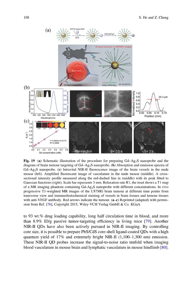
Fig. 19 (a) Schematic illustration of the procedure for preparing Gd–Ag 2 S nanoprobe and the
diagram of brain tumour targeting of Gd–Ag 2 S nanoprobe. (b) Absorption and emission spectra of
Gd–Ag 2 S nanoprobe. (c) Intravital NIR-II fluorescence image of the brain vessels in the nude
mouse (left). Amplified fluorescent image of vasculature in the nude mouse (middle). A crosssectional intensity profile measured along the red-dashed line in (middle) with its peak fitted to
Gaussian functions (right). Scale bar represents 3 mm. Relaxation rate R1, the inset shows a T1 map
of a MR imaging phantom containing Gd-Ag 2 S nanoprobe with different concentrations. In vivo
progressive T1-weighted MR images of the U87MG brain tumour at different time points from
transverse view and immunohistochemical staining of vessels in brain tissues and tumour tissues
with anti-VEGF antibody. Red arrows indicate the tumour. (a–c) Reprinted (adapted) with permission from Ref. [76], Copyright 2015, Wiley-VCH Verlag GmbH & Co. KGaA
108
S. He and Z. Cheng
than 8.9% ID/g passive tumor-targeting efficiency in living mice [79]. Another
NIR-II QDs have also been actively pursued in NIR-II imaging. By controlling
core size, it is possible to prepare PbS/CdS core-shell ligand-coated QDs with a high
quantum yield of 17% and extremely bright NIR-II (1,100–1,300 nm) emission.
These NIR-II QD probes increase the signal-to-noise ratio tenfold when imaging
blood vasculature in mouse brain and lymphatic vasculature in mouse hindlimb [80].
Fig. 19 (a) Schematic illustration of the procedure for preparing Gd–Ag 2 S nanoprobe and the
diagram of brain tumour targeting of Gd–Ag 2 S nanoprobe. (b) Absorption and emission spectra of
Gd–Ag 2 S nanoprobe. (c) Intravital NIR-II fluorescence image of the brain vessels in the nude
mouse (left). Amplified fluorescent image of vasculature in the nude mouse (middle). A crosssectional intensity profile measured along the red-dashed line in (middle) with its peak fitted to
Gaussian functions (right). Scale bar represents 3 mm. Relaxation rate R1, the inset shows a T1 map
of a MR imaging phantom containing Gd-Ag 2 S nanoprobe with different concentrations. In vivo
progressive T1-weighted MR images of the U87MG brain tumour at different time points from
transverse view and immunohistochemical staining of vessels in brain tissues and tumour tissues
with anti-VEGF antibody. Red arrows indicate the tumour. (a–c) Reprinted (adapted) with permission from Ref. [76], Copyright 2015, Wiley-VCH Verlag GmbH & Co. KGaA
108
S. He and Z. Cheng