

34
with fluorescence-based detection methods. This “optical bar code” obtained is simply a combination of fluorescent dyes with different excitation and emission wavelengths and intensities that allow each bead to be independently identified [70].
Nonetheless, additional degrees of freedom are available in the fiber-optic format, which allows an additional number of excitation and emission wavelengths
that can be used. It should be mentioned that the optically bar-coded arrays, can be
decoded in a matter of seconds. This occurs by conventional image processing
software that collects a series of fluorescence images at different excitation and
emission wavelengths and then analyzes the relative intensities of each bead.
Alternatively, preformed oligonucleotides may be added directly to surfaceactivated microspheres [70].
Needless to say, those fluorescence-based assays are more desirable than traditional radiolabeled methods due to their increased safety and experimental versatility. Fluorescence can be incorporated into microarray assays by fluorescent
intercalating dyes, fluorescently labeled targets, or label-less methods employing
fluorescence resonance energy transfer (FRET). Fluorescence-based assays enable
the measurement of multiple wavelengths independently and simultaneously. The
use of multiple fluorophores enables parallel interrogation schemes [70–73].
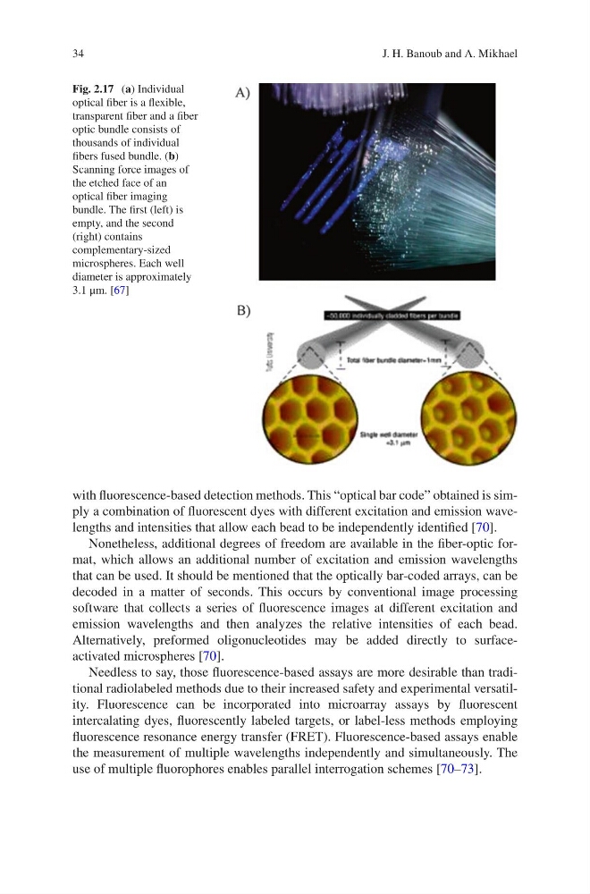
Fig. 2.17 (a) Individual
optical fiber is a flexible,
transparent fiber and a fiber
optic bundle consists of
thousands of individual
fibers fused bundle. (b)
Scanning force images of
the etched face of an
optical fiber imaging
bundle. The first (left) is
empty, and the second
(right) contains
complementary-sized
microspheres. Each well
diameter is approximately
3.1 μm. [67]
J. H. Banoub and A. Mikhael
with fluorescence-based detection methods. This “optical bar code” obtained is simply a combination of fluorescent dyes with different excitation and emission wavelengths and intensities that allow each bead to be independently identified [70].
Nonetheless, additional degrees of freedom are available in the fiber-optic format, which allows an additional number of excitation and emission wavelengths
that can be used. It should be mentioned that the optically bar-coded arrays, can be
decoded in a matter of seconds. This occurs by conventional image processing
software that collects a series of fluorescence images at different excitation and
emission wavelengths and then analyzes the relative intensities of each bead.
Alternatively, preformed oligonucleotides may be added directly to surfaceactivated microspheres [70].
Needless to say, those fluorescence-based assays are more desirable than traditional radiolabeled methods due to their increased safety and experimental versatility. Fluorescence can be incorporated into microarray assays by fluorescent
intercalating dyes, fluorescently labeled targets, or label-less methods employing
fluorescence resonance energy transfer (FRET). Fluorescence-based assays enable
the measurement of multiple wavelengths independently and simultaneously. The
use of multiple fluorophores enables parallel interrogation schemes [70–73].
Fig. 2.17 (a) Individual
optical fiber is a flexible,
transparent fiber and a fiber
optic bundle consists of
thousands of individual
fibers fused bundle. (b)
Scanning force images of
the etched face of an
optical fiber imaging
bundle. The first (left) is
empty, and the second
(right) contains
complementary-sized
microspheres. Each well
diameter is approximately
3.1 μm. [67]
J. H. Banoub and A. Mikhael