

114
7.5 Sample Preparation for IMS
IMS is highly dependent on the quality of sample preparation. Many factors must be
taken into account when preparing a sample, including tissue stabilization or preservation, embedding material, the washing procedure, matrix choice, and matrix
application method [34]. An example workflow consists of tissue sectioning, an
optional washing procedure, matrix application, and an optional recrystallization
procedure (Fig. 7.4). The primary goal of any sample preparation is to minimize
analyte delocalization and degradation while maximizing sensitivity [35]. Sample
preparation protocols are typically developed for specific tissue types and analytes
of interest [36–40], although common methods and protocols can be established.
Sample preparation of formalin fixed tissue [41] begins by sectioning to
10–12 μm thickness and thaw mounting onto a conductive microscope slide
(Fig. 7.4a). Fixation, offering the most accurate morphological tissue representation, is performed by incubating a harvested tissue in a paraformaldehyde solution
for up to 7 days. Intramolecular crosslinks are formed between proteins during this
incubation. The fixed tissue is then submerged in paraffin wax, and is termed formalin fixed paraffin embedded (FFPE) tissue. While this method allows ambient storage, the fixative can induce unwanted issues [42]. These can compromise analysis
of metabolites, lipids, and intact proteins. However, some studies observe limited
MS effects after paraffin removal or alternative procedures [43–45]. For proteins,
developed protocols utilize proteolytic digestion of FFPE tissues and subsequent
peptide analysis [37, 43, 45, 46]. Steps within these protocols allow more efficient
enzyme digestion of proteins. Fresh frozen tissues are preferred for IMS [34].
However, freezing induces morphological changes and other artifacts to a tissue.
Histological stains of fresh frozen tissue sections are typically inferior to those of
FFPE sections.
For fresh frozen tissue, the preserved tissue is cryosectioned prior to thaw mounting onto the slide. Optimal cutting temperature (OCT) compound may be used to
improve section quality. However, OCT is a polymer that introduces high intensity
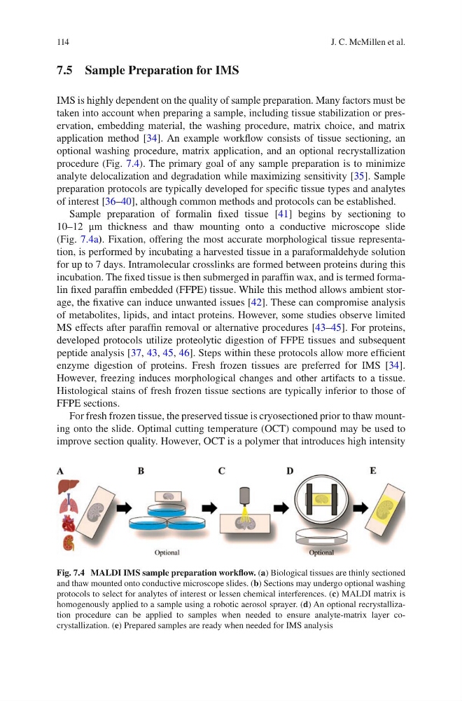
Fig. 7.4 MALDI IMS sample preparation workflow. (a) Biological tissues are thinly sectioned
and thaw mounted onto conductive microscope slides. (b) Sections may undergo optional washing
protocols to select for analytes of interest or lessen chemical interferences. (c) MALDI matrix is
homogenously applied to a sample using a robotic aerosol sprayer. (d) An optional recrystallization procedure can be applied to samples when needed to ensure analyte-matrix layer cocrystallization. (e) Prepared samples are ready when needed for IMS analysis
J. C. McMillen et al.
7.5 Sample Preparation for IMS
IMS is highly dependent on the quality of sample preparation. Many factors must be
taken into account when preparing a sample, including tissue stabilization or preservation, embedding material, the washing procedure, matrix choice, and matrix
application method [34]. An example workflow consists of tissue sectioning, an
optional washing procedure, matrix application, and an optional recrystallization
procedure (Fig. 7.4). The primary goal of any sample preparation is to minimize
analyte delocalization and degradation while maximizing sensitivity [35]. Sample
preparation protocols are typically developed for specific tissue types and analytes
of interest [36–40], although common methods and protocols can be established.
Sample preparation of formalin fixed tissue [41] begins by sectioning to
10–12 μm thickness and thaw mounting onto a conductive microscope slide
(Fig. 7.4a). Fixation, offering the most accurate morphological tissue representation, is performed by incubating a harvested tissue in a paraformaldehyde solution
for up to 7 days. Intramolecular crosslinks are formed between proteins during this
incubation. The fixed tissue is then submerged in paraffin wax, and is termed formalin fixed paraffin embedded (FFPE) tissue. While this method allows ambient storage, the fixative can induce unwanted issues [42]. These can compromise analysis
of metabolites, lipids, and intact proteins. However, some studies observe limited
MS effects after paraffin removal or alternative procedures [43–45]. For proteins,
developed protocols utilize proteolytic digestion of FFPE tissues and subsequent
peptide analysis [37, 43, 45, 46]. Steps within these protocols allow more efficient
enzyme digestion of proteins. Fresh frozen tissues are preferred for IMS [34].
However, freezing induces morphological changes and other artifacts to a tissue.
Histological stains of fresh frozen tissue sections are typically inferior to those of
FFPE sections.
For fresh frozen tissue, the preserved tissue is cryosectioned prior to thaw mounting onto the slide. Optimal cutting temperature (OCT) compound may be used to
improve section quality. However, OCT is a polymer that introduces high intensity
Fig. 7.4 MALDI IMS sample preparation workflow. (a) Biological tissues are thinly sectioned
and thaw mounted onto conductive microscope slides. (b) Sections may undergo optional washing
protocols to select for analytes of interest or lessen chemical interferences. (c) MALDI matrix is
homogenously applied to a sample using a robotic aerosol sprayer. (d) An optional recrystallization procedure can be applied to samples when needed to ensure analyte-matrix layer cocrystallization. (e) Prepared samples are ready when needed for IMS analysis
J. C. McMillen et al.