

132
–3.8
–3.9
–4.0
–4.1
–4.2
BE-O (eV)
–3.20
–3.10
–3.00
Surface strain (%)
defect
Pt
Ni
N
Pt diffusion
to defective sites
–4.15 –4.10 –4.05 –4.00 –3.95 –3.90
BE-O (eV)
1.8
1.5
1.2
0.9
0.6
0.3
0.0
Specific activity (mA/cm
2
)
PtNiN/C
Pt/C
Pt
Pt 2ML Ni 4 N
(a)
(c)
(b)
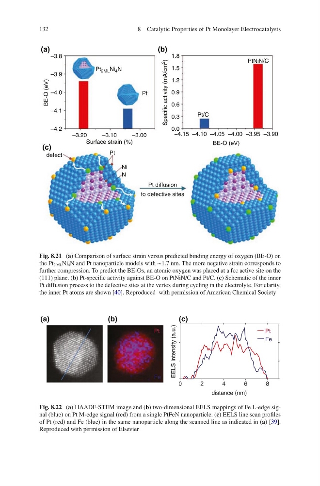
Fig. 8.21 (a) Comparison of surface strain versus predicted binding energy of oxygen (BE-O) on
the Pt 2 ML Ni 4 N and Pt nanoparticle models with ∼1.7 nm. The more negative strain corresponds to
further compression. To predict the BE-Os, an atomic oxygen was placed at a fcc active site on the
(111) plane. (b) Pt-specific activity against BE-O on PtNiN/C and Pt/C. (c) Schematic of the inner
Pt diffusion process to the defective sites at the vertex during cycling in the electrolyte. For clarity,
the inner Pt atoms are shown [40]. Reproduced with permission of American Chemical Society
0
2
4
6
8
distance (nm)
EELS intensity (a.u.)
Pt
Pt
Fe
Fe
(a)
(b)
(c)
Fig. 8.22 (a) HAADF-STEM image and (b) two-dimensional EELS mappings of Fe L-edge signal (blue) on Pt M-edge signal (red) from a single PtFeN nanoparticle. (c) EELS line scan profiles
of Pt (red) and Fe (blue) in the same nanoparticle along the scanned line as indicated in (a) [39].
Reproduced with permission of Elsevier
8 Catalytic Properties of Pt Monolayer Electrocatalysts
–3.8
–3.9
–4.0
–4.1
–4.2
BE-O (eV)
–3.20
–3.10
–3.00
Surface strain (%)
defect
Pt
Ni
N
Pt diffusion
to defective sites
–4.15 –4.10 –4.05 –4.00 –3.95 –3.90
BE-O (eV)
1.8
1.5
1.2
0.9
0.6
0.3
0.0
Specific activity (mA/cm
2
)
PtNiN/C
Pt/C
Pt
Pt 2ML Ni 4 N
(a)
(c)
(b)
Fig. 8.21 (a) Comparison of surface strain versus predicted binding energy of oxygen (BE-O) on
the Pt 2 ML Ni 4 N and Pt nanoparticle models with ∼1.7 nm. The more negative strain corresponds to
further compression. To predict the BE-Os, an atomic oxygen was placed at a fcc active site on the
(111) plane. (b) Pt-specific activity against BE-O on PtNiN/C and Pt/C. (c) Schematic of the inner
Pt diffusion process to the defective sites at the vertex during cycling in the electrolyte. For clarity,
the inner Pt atoms are shown [40]. Reproduced with permission of American Chemical Society
0
2
4
6
8
distance (nm)
EELS intensity (a.u.)
Pt
Pt
Fe
Fe
(a)
(b)
(c)
Fig. 8.22 (a) HAADF-STEM image and (b) two-dimensional EELS mappings of Fe L-edge signal (blue) on Pt M-edge signal (red) from a single PtFeN nanoparticle. (c) EELS line scan profiles
of Pt (red) and Fe (blue) in the same nanoparticle along the scanned line as indicated in (a) [39].
Reproduced with permission of Elsevier
8 Catalytic Properties of Pt Monolayer Electrocatalysts