

460
8 Morphogenesis
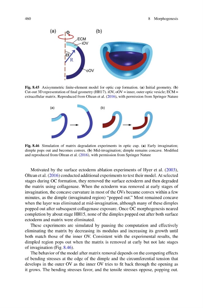
Fig. 8.45 Axisymmetric finite-element model for optic cup formation. (a) Initial geometry. (b)
Cut-out 3D representation of final geometry (HH17). iOV, oOV = inner, outer optic vesicle; ECM =
extracellular matrix. Reproduced from Oltean et al. (2016), with permission from Springer Nature
Fig. 8.46 Simulation of matrix degradation experiments in optic cup. (a) Early invagination;
dimple pops out and becomes convex. (b) Mid-invagination; dimple remains concave. Modified
and reproduced from Oltean et al. (2016), with permission from Springer Nature
Motivated by the surface ectoderm ablation experiments of Hyer et al. (2003),
Oltean et al. (2016) conducted additional experiments to test their model. At selected
stages during OC formation, they removed the surface ectoderm and then degraded
the matrix using collagenase. When the ectoderm was removed at early stages of
invagination, the concave curvature in most of the OVs became convex within a few
minutes, as the dimple (invaginated region) “popped out.” Most remained concave
when the layer was eliminated at mid-invagination, although many of these dimples
popped out after subsequent collagenase exposure. Once OC morphogenesis neared
completion by about stage HH15, none of the dimples popped out after both surface
ectoderm and matrix were eliminated.
These experiments are simulated by pausing the computation and effectively
eliminating the matrix by decreasing its modulus and increasing its growth until
both match those of the inner OV. Consistent with the experimental results, the
dimpled region pops out when the matrix is removed at early but not late stages
of invagination (Fig. 8.46).
The behavior of the model after matrix removal depends on the competing effects
of bending stresses at the edge of the dimple and the circumferential tension that
develops in the outer OV as the inner OV tries to fit back through the opening as
it grows. The bending stresses favor, and the tensile stresses oppose, popping out.
8 Morphogenesis
Fig. 8.45 Axisymmetric finite-element model for optic cup formation. (a) Initial geometry. (b)
Cut-out 3D representation of final geometry (HH17). iOV, oOV = inner, outer optic vesicle; ECM =
extracellular matrix. Reproduced from Oltean et al. (2016), with permission from Springer Nature
Fig. 8.46 Simulation of matrix degradation experiments in optic cup. (a) Early invagination;
dimple pops out and becomes convex. (b) Mid-invagination; dimple remains concave. Modified
and reproduced from Oltean et al. (2016), with permission from Springer Nature
Motivated by the surface ectoderm ablation experiments of Hyer et al. (2003),
Oltean et al. (2016) conducted additional experiments to test their model. At selected
stages during OC formation, they removed the surface ectoderm and then degraded
the matrix using collagenase. When the ectoderm was removed at early stages of
invagination, the concave curvature in most of the OVs became convex within a few
minutes, as the dimple (invaginated region) “popped out.” Most remained concave
when the layer was eliminated at mid-invagination, although many of these dimples
popped out after subsequent collagenase exposure. Once OC morphogenesis neared
completion by about stage HH15, none of the dimples popped out after both surface
ectoderm and matrix were eliminated.
These experiments are simulated by pausing the computation and effectively
eliminating the matrix by decreasing its modulus and increasing its growth until
both match those of the inner OV. Consistent with the experimental results, the
dimpled region pops out when the matrix is removed at early but not late stages
of invagination (Fig. 8.46).
The behavior of the model after matrix removal depends on the competing effects
of bending stresses at the edge of the dimple and the circumferential tension that
develops in the outer OV as the inner OV tries to fit back through the opening as
it grows. The bending stresses favor, and the tensile stresses oppose, popping out.