

8.1 Cellular Mechanisms of Epithelial Morphogenesis
403
8.1.1 Structure of Epithelia
Embryonic epithelia consist of one or more layers of cells that often have hexagonal
shapes (Figs. 8.1 and 8.3). The basal surface is attached to a basement membrane
composed of extracellular matrix, whereas the apical surface faces open space such
as a lumen. The component cells are attached to each other by cell-cell adhesion
molecules (e.g., cadherins) and to the basement membrane by cell-matrix adhesion
molecules (e.g., integrins) (Alberts et al. 2014).
The adhesions linking cells are organized as circumferential rings called tight
junctions near the cell apex and adherens junctions below (Fig. 8.1). Tight
junctions serve as barriers to transport across the epithelium. Adherens junctions
transmit forces to the actin cytoskeleton and to an associated actomyosin belt around
the cell. During morphogenesis, adherens junctions transmit forces between cells,
enabling epithelia to undergo large-scale deformations (Martin et al. 2010). Another
type of junction, called a gap junction, is a pore that allows the passage of small
molecules between cells, including morphogens.
8.1.2 Stretching Morphogenesis
Epithelial stretching involves planar changes in size and shape. At the cell level,
these changes can be produced in various ways, including the following (Davies
2005; Keller 2006; Taber 2014a):
1. Cells divide or die, hypertrophy or atrophy (Fig. 8.2a, b). Examples: expansion of
the heart tube (Soufan et al. 2006); closure of the neural tube by lateral extension
of adjacent ectoderm (Colas and Schoenwolf 2001) (see Fig. 1.4).
2. New cells are added by the process of mesenchymal-epithelial transition
(Fig. 8.2c), or old cells leave by epithelial-mesenchymal transition. Examples:
mesenchymal cells that become epithelial cells to form the blastula (Gilbert
2010) and primitive heart fields (Kirby 2007) (see Sect. 1.4.2).
Tight junction
Adherens junction/
actomyosin fibers
Apex
Base
Contract
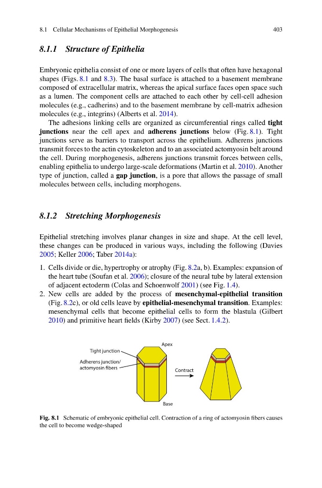
Fig. 8.1 Schematic of embryonic epithelial cell. Contraction of a ring of actomyosin fibers causes
the cell to become wedge-shaped
403
8.1.1 Structure of Epithelia
Embryonic epithelia consist of one or more layers of cells that often have hexagonal
shapes (Figs. 8.1 and 8.3). The basal surface is attached to a basement membrane
composed of extracellular matrix, whereas the apical surface faces open space such
as a lumen. The component cells are attached to each other by cell-cell adhesion
molecules (e.g., cadherins) and to the basement membrane by cell-matrix adhesion
molecules (e.g., integrins) (Alberts et al. 2014).
The adhesions linking cells are organized as circumferential rings called tight
junctions near the cell apex and adherens junctions below (Fig. 8.1). Tight
junctions serve as barriers to transport across the epithelium. Adherens junctions
transmit forces to the actin cytoskeleton and to an associated actomyosin belt around
the cell. During morphogenesis, adherens junctions transmit forces between cells,
enabling epithelia to undergo large-scale deformations (Martin et al. 2010). Another
type of junction, called a gap junction, is a pore that allows the passage of small
molecules between cells, including morphogens.
8.1.2 Stretching Morphogenesis
Epithelial stretching involves planar changes in size and shape. At the cell level,
these changes can be produced in various ways, including the following (Davies
2005; Keller 2006; Taber 2014a):
1. Cells divide or die, hypertrophy or atrophy (Fig. 8.2a, b). Examples: expansion of
the heart tube (Soufan et al. 2006); closure of the neural tube by lateral extension
of adjacent ectoderm (Colas and Schoenwolf 2001) (see Fig. 1.4).
2. New cells are added by the process of mesenchymal-epithelial transition
(Fig. 8.2c), or old cells leave by epithelial-mesenchymal transition. Examples:
mesenchymal cells that become epithelial cells to form the blastula (Gilbert
2010) and primitive heart fields (Kirby 2007) (see Sect. 1.4.2).
Tight junction
Adherens junction/
actomyosin fibers
Apex
Base
Contract
Fig. 8.1 Schematic of embryonic epithelial cell. Contraction of a ring of actomyosin fibers causes
the cell to become wedge-shaped