

1.4 Developmental Biology Background
9
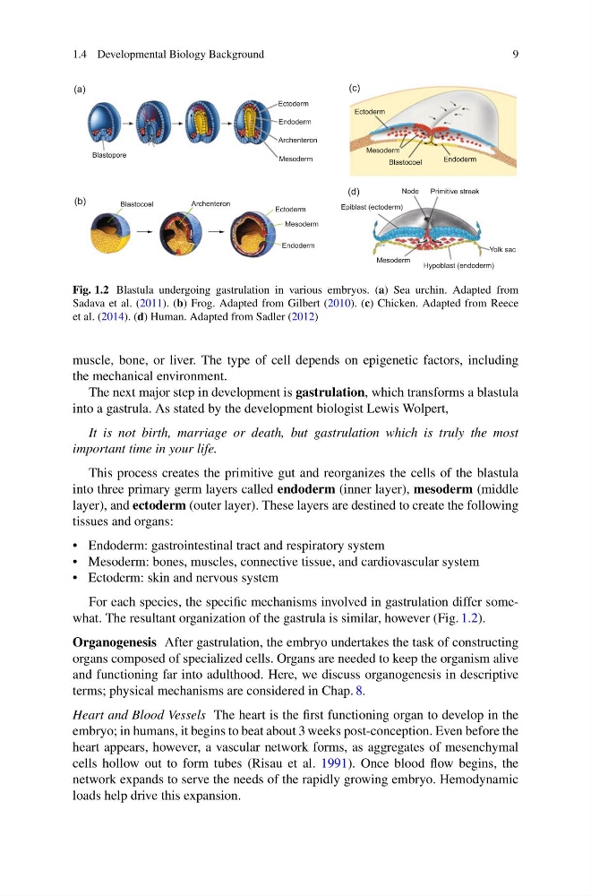
Fig. 1.2 Blastula undergoing gastrulation in various embryos. (a) Sea urchin. Adapted from
Sadava et al. (2011). (b) Frog. Adapted from Gilbert (2010). (c) Chicken. Adapted from Reece
et al. (2014). (d) Human. Adapted from Sadler (2012)
muscle, bone, or liver. The type of cell depends on epigenetic factors, including
the mechanical environment.
The next major step in development is gastrulation, which transforms a blastula
into a gastrula. As stated by the development biologist Lewis Wolpert,
It is not birth, marriage or death, but gastrulation which is truly the most
important time in your life.
This process creates the primitive gut and reorganizes the cells of the blastula
into three primary germ layers called endoderm (inner layer), mesoderm (middle
layer), and ectoderm (outer layer). These layers are destined to create the following
tissues and organs:
• Endoderm: gastrointestinal tract and respiratory system
• Mesoderm: bones, muscles, connective tissue, and cardiovascular system
• Ectoderm: skin and nervous system
For each species, the specific mechanisms involved in gastrulation differ somewhat. The resultant organization of the gastrula is similar, however (Fig. 1.2).
Organogenesis After gastrulation, the embryo undertakes the task of constructing
organs composed of specialized cells. Organs are needed to keep the organism alive
and functioning far into adulthood. Here, we discuss organogenesis in descriptive
terms; physical mechanisms are considered in Chap. 8.
Heart and Blood Vessels The heart is the first functioning organ to develop in the
embryo; in humans, it begins to beat about 3 weeks post-conception. Even before the
heart appears, however, a vascular network forms, as aggregates of mesenchymal
cells hollow out to form tubes (Risau et al. 1991). Once blood flow begins, the
network expands to serve the needs of the rapidly growing embryo. Hemodynamic
loads help drive this expansion.
9
Fig. 1.2 Blastula undergoing gastrulation in various embryos. (a) Sea urchin. Adapted from
Sadava et al. (2011). (b) Frog. Adapted from Gilbert (2010). (c) Chicken. Adapted from Reece
et al. (2014). (d) Human. Adapted from Sadler (2012)
muscle, bone, or liver. The type of cell depends on epigenetic factors, including
the mechanical environment.
The next major step in development is gastrulation, which transforms a blastula
into a gastrula. As stated by the development biologist Lewis Wolpert,
It is not birth, marriage or death, but gastrulation which is truly the most
important time in your life.
This process creates the primitive gut and reorganizes the cells of the blastula
into three primary germ layers called endoderm (inner layer), mesoderm (middle
layer), and ectoderm (outer layer). These layers are destined to create the following
tissues and organs:
• Endoderm: gastrointestinal tract and respiratory system
• Mesoderm: bones, muscles, connective tissue, and cardiovascular system
• Ectoderm: skin and nervous system
For each species, the specific mechanisms involved in gastrulation differ somewhat. The resultant organization of the gastrula is similar, however (Fig. 1.2).
Organogenesis After gastrulation, the embryo undertakes the task of constructing
organs composed of specialized cells. Organs are needed to keep the organism alive
and functioning far into adulthood. Here, we discuss organogenesis in descriptive
terms; physical mechanisms are considered in Chap. 8.
Heart and Blood Vessels The heart is the first functioning organ to develop in the
embryo; in humans, it begins to beat about 3 weeks post-conception. Even before the
heart appears, however, a vascular network forms, as aggregates of mesenchymal
cells hollow out to form tubes (Risau et al. 1991). Once blood flow begins, the
network expands to serve the needs of the rapidly growing embryo. Hemodynamic
loads help drive this expansion.