

220
5 Contraction
Active Force
Time
Active Force
Length
Velocity
Force
(a)
(b)
(c)
tetanus
twitch
clamp
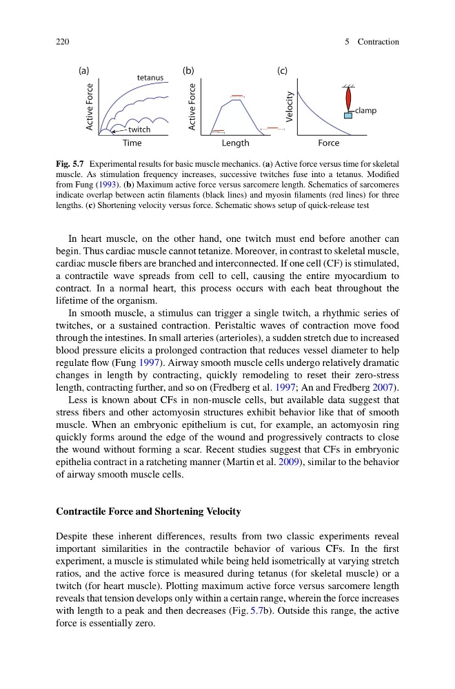
Fig. 5.7 Experimental results for basic muscle mechanics. (a) Active force versus time for skeletal
muscle. As stimulation frequency increases, successive twitches fuse into a tetanus. Modified
from Fung (1993). (b) Maximum active force versus sarcomere length. Schematics of sarcomeres
indicate overlap between actin filaments (black lines) and myosin filaments (red lines) for three
lengths. (c) Shortening velocity versus force. Schematic shows setup of quick-release test
In heart muscle, on the other hand, one twitch must end before another can
begin. Thus cardiac muscle cannot tetanize. Moreover, in contrast to skeletal muscle,
cardiac muscle fibers are branched and interconnected. If one cell (CF) is stimulated,
a contractile wave spreads from cell to cell, causing the entire myocardium to
contract. In a normal heart, this process occurs with each beat throughout the
lifetime of the organism.
In smooth muscle, a stimulus can trigger a single twitch, a rhythmic series of
twitches, or a sustained contraction. Peristaltic waves of contraction move food
through the intestines. In small arteries (arterioles), a sudden stretch due to increased
blood pressure elicits a prolonged contraction that reduces vessel diameter to help
regulate flow (Fung 1997). Airway smooth muscle cells undergo relatively dramatic
changes in length by contracting, quickly remodeling to reset their zero-stress
length, contracting further, and so on (Fredberg et al. 1997; An and Fredberg 2007).
Less is known about CFs in non-muscle cells, but available data suggest that
stress fibers and other actomyosin structures exhibit behavior like that of smooth
muscle. When an embryonic epithelium is cut, for example, an actomyosin ring
quickly forms around the edge of the wound and progressively contracts to close
the wound without forming a scar. Recent studies suggest that CFs in embryonic
epithelia contract in a ratcheting manner (Martin et al. 2009), similar to the behavior
of airway smooth muscle cells.
Contractile Force and Shortening Velocity
Despite these inherent differences, results from two classic experiments reveal
important similarities in the contractile behavior of various CFs. In the first
experiment, a muscle is stimulated while being held isometrically at varying stretch
ratios, and the active force is measured during tetanus (for skeletal muscle) or a
twitch (for heart muscle). Plotting maximum active force versus sarcomere length
reveals that tension develops only within a certain range, wherein the force increases
with length to a peak and then decreases (Fig. 5.7b). Outside this range, the active
force is essentially zero.
5 Contraction
Active Force
Time
Active Force
Length
Velocity
Force
(a)
(b)
(c)
tetanus
twitch
clamp
Fig. 5.7 Experimental results for basic muscle mechanics. (a) Active force versus time for skeletal
muscle. As stimulation frequency increases, successive twitches fuse into a tetanus. Modified
from Fung (1993). (b) Maximum active force versus sarcomere length. Schematics of sarcomeres
indicate overlap between actin filaments (black lines) and myosin filaments (red lines) for three
lengths. (c) Shortening velocity versus force. Schematic shows setup of quick-release test
In heart muscle, on the other hand, one twitch must end before another can
begin. Thus cardiac muscle cannot tetanize. Moreover, in contrast to skeletal muscle,
cardiac muscle fibers are branched and interconnected. If one cell (CF) is stimulated,
a contractile wave spreads from cell to cell, causing the entire myocardium to
contract. In a normal heart, this process occurs with each beat throughout the
lifetime of the organism.
In smooth muscle, a stimulus can trigger a single twitch, a rhythmic series of
twitches, or a sustained contraction. Peristaltic waves of contraction move food
through the intestines. In small arteries (arterioles), a sudden stretch due to increased
blood pressure elicits a prolonged contraction that reduces vessel diameter to help
regulate flow (Fung 1997). Airway smooth muscle cells undergo relatively dramatic
changes in length by contracting, quickly remodeling to reset their zero-stress
length, contracting further, and so on (Fredberg et al. 1997; An and Fredberg 2007).
Less is known about CFs in non-muscle cells, but available data suggest that
stress fibers and other actomyosin structures exhibit behavior like that of smooth
muscle. When an embryonic epithelium is cut, for example, an actomyosin ring
quickly forms around the edge of the wound and progressively contracts to close
the wound without forming a scar. Recent studies suggest that CFs in embryonic
epithelia contract in a ratcheting manner (Martin et al. 2009), similar to the behavior
of airway smooth muscle cells.
Contractile Force and Shortening Velocity
Despite these inherent differences, results from two classic experiments reveal
important similarities in the contractile behavior of various CFs. In the first
experiment, a muscle is stimulated while being held isometrically at varying stretch
ratios, and the active force is measured during tetanus (for skeletal muscle) or a
twitch (for heart muscle). Plotting maximum active force versus sarcomere length
reveals that tension develops only within a certain range, wherein the force increases
with length to a peak and then decreases (Fig. 5.7b). Outside this range, the active
force is essentially zero.