

achieved due to insufficient interaction with the SPE sorbent [53]. This observation
might be extrapolated also to sugar conjugates from other drugs.
As an alternative pretreatment, a modified QuEChERS approach for extracting
parent drugs and their metabolites from plants was used, allowing skipping the SPE
step and still providing good limits of detection for both parent drugs and metabolites [35, 45, 54, 64]. One way to improve extraction by an increased cell-rupturing
effect, setting free also drug and drug-metabolite molecules within the plant cells, is
the use of methods involving matrix solid phase dispersion (MSPD) [65] or sea sand
disruption [65]. MSPD is based on the blending of a sample with an abrasive solid
material [66]. It permits simultaneous disruption of the sample architecture and
extraction of complex biological samples, thereby improving extraction efficiency.
The workflow discussed above, although most widely used, involving either
extraction of the whole plant or dissection of plant parts with subsequent analysis
of the compounds of interest does only allow a rough estimation on the location of
parent drugs and their metabolites within the plant. An alternative approach,
enabling direct investigations on the spatial distribution of the drug and its metabolites within plant parts, is mass spectrometry imaging. Hereby the sample of interest
(plant part) is examined, after appropriate sample preparation, by techniques such as
desorption electrospray ionization (DESI) MS or matrix-assisted laser desorption
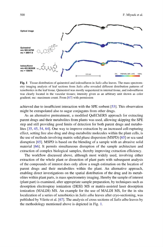
ionization (MALDI) MS. An example for the use of MALDI MS, for the in situ
localization of a series of xenobiotics in Salix alba leaves after cryo-sectioning, was
published by Vilette et al. [67]. The analysis of cross sections of Salix alba leaves by
the methodology mentioned above is depicted in Fig. 1.
Fig. 1 Tissue distribution of quinestrol and iodosulfuron in Salix alba leaves. The mass spectrometry imaging analysis of leaf sections from Salix alba revealed different distribution patterns of
xenobiotics in the leaf tissue. Quinestrol was mostly sequestered in internal tissue, and iodosulfuron
was clearly located in the vascular tissues; intensity given as an arbitrary unit shown as color
gradient; mc: maximum count. From [67] with permission
500
F. Mlynek et al.
might be extrapolated also to sugar conjugates from other drugs.
As an alternative pretreatment, a modified QuEChERS approach for extracting
parent drugs and their metabolites from plants was used, allowing skipping the SPE
step and still providing good limits of detection for both parent drugs and metabolites [35, 45, 54, 64]. One way to improve extraction by an increased cell-rupturing
effect, setting free also drug and drug-metabolite molecules within the plant cells, is
the use of methods involving matrix solid phase dispersion (MSPD) [65] or sea sand
disruption [65]. MSPD is based on the blending of a sample with an abrasive solid
material [66]. It permits simultaneous disruption of the sample architecture and
extraction of complex biological samples, thereby improving extraction efficiency.
The workflow discussed above, although most widely used, involving either
extraction of the whole plant or dissection of plant parts with subsequent analysis
of the compounds of interest does only allow a rough estimation on the location of
parent drugs and their metabolites within the plant. An alternative approach,
enabling direct investigations on the spatial distribution of the drug and its metabolites within plant parts, is mass spectrometry imaging. Hereby the sample of interest
(plant part) is examined, after appropriate sample preparation, by techniques such as
desorption electrospray ionization (DESI) MS or matrix-assisted laser desorption
ionization (MALDI) MS. An example for the use of MALDI MS, for the in situ
localization of a series of xenobiotics in Salix alba leaves after cryo-sectioning, was
published by Vilette et al. [67]. The analysis of cross sections of Salix alba leaves by
the methodology mentioned above is depicted in Fig. 1.
Fig. 1 Tissue distribution of quinestrol and iodosulfuron in Salix alba leaves. The mass spectrometry imaging analysis of leaf sections from Salix alba revealed different distribution patterns of
xenobiotics in the leaf tissue. Quinestrol was mostly sequestered in internal tissue, and iodosulfuron
was clearly located in the vascular tissues; intensity given as an arbitrary unit shown as color
gradient; mc: maximum count. From [67] with permission
500
F. Mlynek et al.