

addition of the internal standard ε-HCH and 8–12 h extraction in a Soxhlet apparatus
with n-hexane. The detailed procedure including the clean-up and fractionation steps
can be found in Kallenborn et al. (1991).
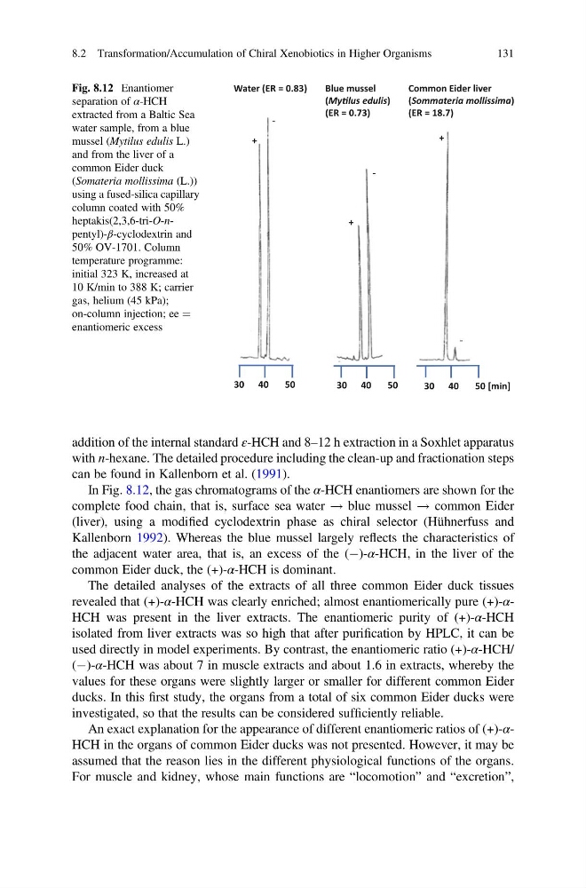
In Fig. 8.12, the gas chromatograms of the α-HCH enantiomers are shown for the
complete food chain, that is, surface sea water ! blue mussel ! common Eider
(liver), using a modified cyclodextrin phase as chiral selector (Hühnerfuss and
Kallenborn 1992). Whereas the blue mussel largely reflects the characteristics of
the adjacent water area, that is, an excess of the (À)-α-HCH, in the liver of the
common Eider duck, the (+)-α-HCH is dominant.
The detailed analyses of the extracts of all three common Eider duck tissues
revealed that (+)-α-HCH was clearly enriched; almost enantiomerically pure (+)-αHCH was present in the liver extracts. The enantiomeric purity of (+)-α-HCH
isolated from liver extracts was so high that after purification by HPLC, it can be
used directly in model experiments. By contrast, the enantiomeric ratio (+)-α-HCH/
(À)-α-HCH was about 7 in muscle extracts and about 1.6 in extracts, whereby the
values for these organs were slightly larger or smaller for different common Eider
ducks. In this first study, the organs from a total of six common Eider ducks were
investigated, so that the results can be considered sufficiently reliable.
An exact explanation for the appearance of different enantiomeric ratios of (+)-αHCH in the organs of common Eider ducks was not presented. However, it may be
assumed that the reason lies in the different physiological functions of the organs.
For muscle and kidney, whose main functions are “locomotion” and “excretion”,
Water (ER = 0.83)
Blue mussel
(MyƟlus edulis)
(ER = 0.73)
Common Eider liver
(Sommateria mollissima)
(ER = 18.7)
+
‐
+
+
‐
‐
30 40
50
30 40
50
30 40
50 [min]
Fig. 8.12 Enantiomer
separation of α-HCH
extracted from a Baltic Sea
water sample, from a blue
mussel (Mytilus edulis L.)
and from the liver of a
common Eider duck
(Somateria mollissima (L.))
using a fused-silica capillary
column coated with 50%
heptakis(2,3,6-tri-O-npentyl)-β-cyclodextrin and
50% OV-1701. Column
temperature programme:
initial 323 K, increased at
10 K/min to 388 K; carrier
gas, helium (45 kPa);
on-column injection; ee ¼
enantiomeric excess
8.2 Transformation/Accumulation of Chiral Xenobiotics in Higher Organisms
131
with n-hexane. The detailed procedure including the clean-up and fractionation steps
can be found in Kallenborn et al. (1991).
In Fig. 8.12, the gas chromatograms of the α-HCH enantiomers are shown for the
complete food chain, that is, surface sea water ! blue mussel ! common Eider
(liver), using a modified cyclodextrin phase as chiral selector (Hühnerfuss and
Kallenborn 1992). Whereas the blue mussel largely reflects the characteristics of
the adjacent water area, that is, an excess of the (À)-α-HCH, in the liver of the
common Eider duck, the (+)-α-HCH is dominant.
The detailed analyses of the extracts of all three common Eider duck tissues
revealed that (+)-α-HCH was clearly enriched; almost enantiomerically pure (+)-αHCH was present in the liver extracts. The enantiomeric purity of (+)-α-HCH
isolated from liver extracts was so high that after purification by HPLC, it can be
used directly in model experiments. By contrast, the enantiomeric ratio (+)-α-HCH/
(À)-α-HCH was about 7 in muscle extracts and about 1.6 in extracts, whereby the
values for these organs were slightly larger or smaller for different common Eider
ducks. In this first study, the organs from a total of six common Eider ducks were
investigated, so that the results can be considered sufficiently reliable.
An exact explanation for the appearance of different enantiomeric ratios of (+)-αHCH in the organs of common Eider ducks was not presented. However, it may be
assumed that the reason lies in the different physiological functions of the organs.
For muscle and kidney, whose main functions are “locomotion” and “excretion”,
Water (ER = 0.83)
Blue mussel
(MyƟlus edulis)
(ER = 0.73)
Common Eider liver
(Sommateria mollissima)
(ER = 18.7)
+
‐
+
+
‐
‐
30 40
50
30 40
50
30 40
50 [min]
Fig. 8.12 Enantiomer
separation of α-HCH
extracted from a Baltic Sea
water sample, from a blue
mussel (Mytilus edulis L.)
and from the liver of a
common Eider duck
(Somateria mollissima (L.))
using a fused-silica capillary
column coated with 50%
heptakis(2,3,6-tri-O-npentyl)-β-cyclodextrin and
50% OV-1701. Column
temperature programme:
initial 323 K, increased at
10 K/min to 388 K; carrier
gas, helium (45 kPa);
on-column injection; ee ¼
enantiomeric excess
8.2 Transformation/Accumulation of Chiral Xenobiotics in Higher Organisms
131