

112
A. Bagaria and S. Ramakumar
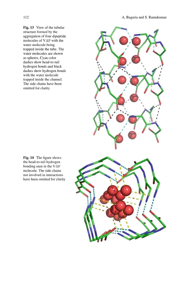
Fig. 13 View of the tubular
structure formed by the
aggregation of four dipeptide
molecules of VF with the
water molecule being
trapped inside the tube. The
water molecules are shown
as spheres. Cyan color
dashes show head-to-tail
hydrogen bonds and black
dashes show hydrogen bonds
with the water molecule
trapped inside the channel.
The side chains have been
omitted for clarity
Fig. 14 The figure shows
the head-to-tail hydrogen
bonding seen in the VF
molecule. The side chains
not involved in interactions
have been omitted for clarity
A. Bagaria and S. Ramakumar
Fig. 13 View of the tubular
structure formed by the
aggregation of four dipeptide
molecules of VF with the
water molecule being
trapped inside the tube. The
water molecules are shown
as spheres. Cyan color
dashes show head-to-tail
hydrogen bonds and black
dashes show hydrogen bonds
with the water molecule
trapped inside the channel.
The side chains have been
omitted for clarity
Fig. 14 The figure shows
the head-to-tail hydrogen
bonding seen in the VF
molecule. The side chains
not involved in interactions
have been omitted for clarity