

102
A. Bagaria and S. Ramakumar
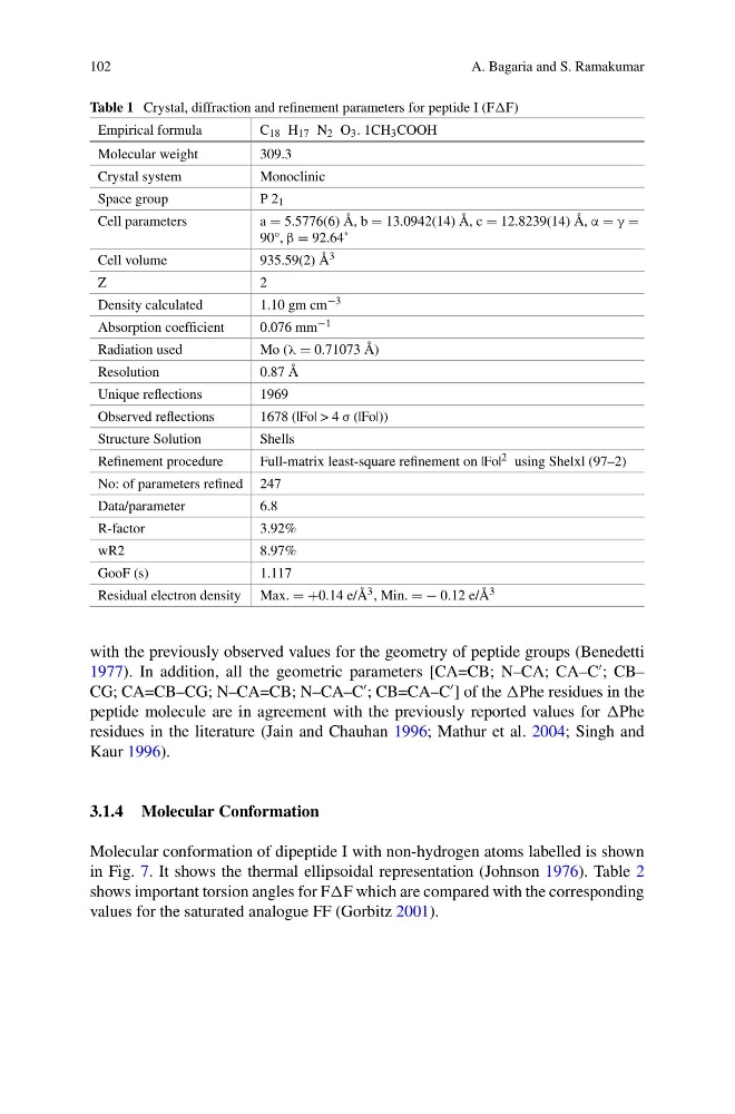
Table 1 Crystal, diffraction and refinement parameters for peptide I (FF)
Empirical formula
C 18 H 17 N 2 O 3 . 1CH 3 COOH
Molecular weight
309.3
Crystal system
Monoclinic
Space group
P 2 1
Cell parameters
a = 5.5776(6) Å, b = 13.0942(14) Å, c = 12.8239(14) Å, α = γ =
90°, β = 92.64˚
Cell volume
935.59(2) Å 3
Z
2
Density calculated
1.10 gm cm −3
Absorption coefficient
0.076 mm −1
Radiation used
Mo (λ = 0.71073 Å)
Resolution
0.87 Å
Unique reflections
1969
Observed reflections
1678 (|Fo| > 4 σ (|Fo|))
Structure Solution
Shells
Refinement procedure
Full-matrix least-square refinement on |Fo| 2 using Shelxl (97–2)
No: of parameters refined 247
Data/parameter
6.8
R-factor
3.92%
wR2
8.97%
GooF (s)
1.117
Residual electron density Max. = +0.14 e/Å 3 , Min. = − 0.12 e/Å 3
with the previously observed values for the geometry of peptide groups (Benedetti
1977). In addition, all the geometric parameters [CA=CB; N–CA; CA–C
; CB–
CG; CA=CB–CG; N–CA=CB; N–CA–C
; CB=CA–C
] of the Phe residues in the
peptide molecule are in agreement with the previously reported values for Phe
residues in the literature (Jain and Chauhan 1996; Mathur et al. 2004; Singh and
Kaur 1996).
3.1.4 Molecular Conformation
Molecular conformation of dipeptide I with non-hydrogen atoms labelled is shown
in Fig. 7. It shows the thermal ellipsoidal representation (Johnson 1976). Table 2
shows important torsion angles for FF which are compared with the corresponding
values for the saturated analogue FF (Gorbitz 2001).
A. Bagaria and S. Ramakumar
Table 1 Crystal, diffraction and refinement parameters for peptide I (FF)
Empirical formula
C 18 H 17 N 2 O 3 . 1CH 3 COOH
Molecular weight
309.3
Crystal system
Monoclinic
Space group
P 2 1
Cell parameters
a = 5.5776(6) Å, b = 13.0942(14) Å, c = 12.8239(14) Å, α = γ =
90°, β = 92.64˚
Cell volume
935.59(2) Å 3
Z
2
Density calculated
1.10 gm cm −3
Absorption coefficient
0.076 mm −1
Radiation used
Mo (λ = 0.71073 Å)
Resolution
0.87 Å
Unique reflections
1969
Observed reflections
1678 (|Fo| > 4 σ (|Fo|))
Structure Solution
Shells
Refinement procedure
Full-matrix least-square refinement on |Fo| 2 using Shelxl (97–2)
No: of parameters refined 247
Data/parameter
6.8
R-factor
3.92%
wR2
8.97%
GooF (s)
1.117
Residual electron density Max. = +0.14 e/Å 3 , Min. = − 0.12 e/Å 3
with the previously observed values for the geometry of peptide groups (Benedetti
1977). In addition, all the geometric parameters [CA=CB; N–CA; CA–C
; CB–
CG; CA=CB–CG; N–CA=CB; N–CA–C
; CB=CA–C
] of the Phe residues in the
peptide molecule are in agreement with the previously reported values for Phe
residues in the literature (Jain and Chauhan 1996; Mathur et al. 2004; Singh and
Kaur 1996).
3.1.4 Molecular Conformation
Molecular conformation of dipeptide I with non-hydrogen atoms labelled is shown
in Fig. 7. It shows the thermal ellipsoidal representation (Johnson 1976). Table 2
shows important torsion angles for FF which are compared with the corresponding
values for the saturated analogue FF (Gorbitz 2001).