

110
Algae
termed ocellus. It is situated toward the left side of the ventral surface of the cell. It consists of a
refractile structure termed hyalosome, thought to act as a lens, subtended by a domed pigmented
part, divided into two sections, a retinoid and a pigmented cup. Between the lens and the retinoid is
a chamber representing an invagination of the cell covering, which is lined by the cell membrane,
and allows the contact of the ocellus with the external medium. The pigmented cup wrapping the
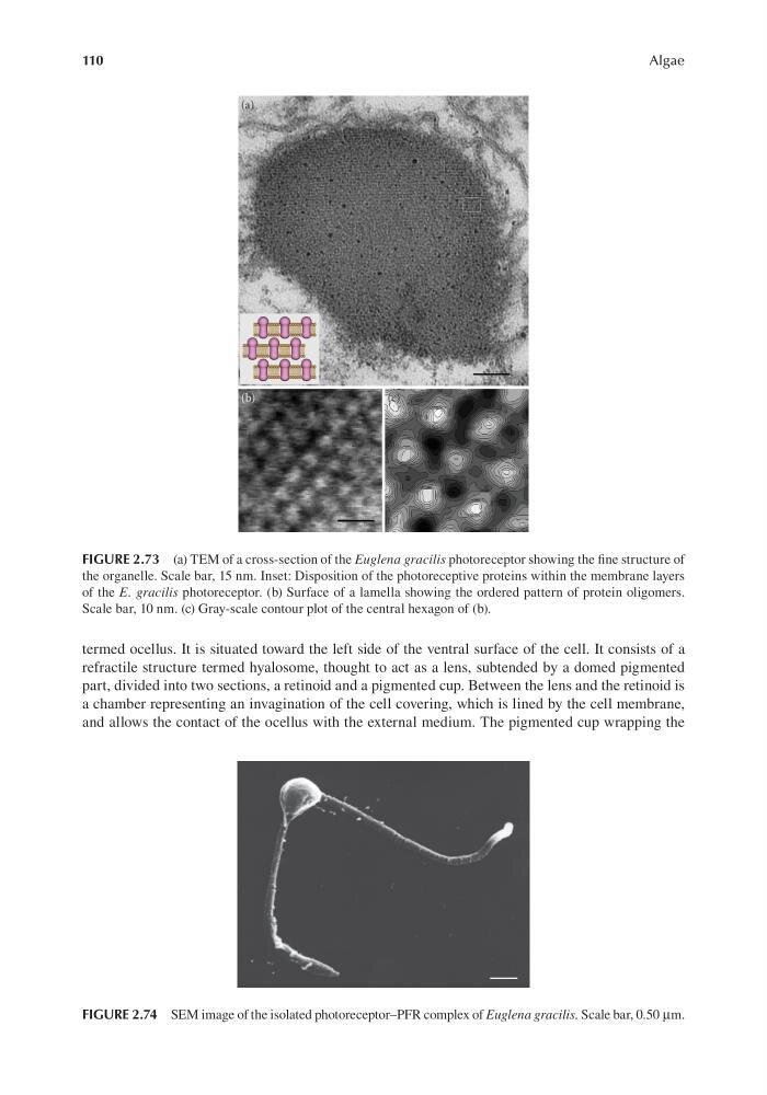
FIGURE 2.73 (a) TEM of a cross-section of the Euglena gracilis photoreceptor showing the fine structure of
the organelle. Scale bar, 15 nm. Inset: Disposition of the photoreceptive proteins within the membrane layers
of the E. gracilis photoreceptor. (b) Surface of a lamella showing the ordered pattern of protein oligomers.
Scale bar, 10 nm. (c) Gray-scale contour plot of the central hexagon of (b).
FIGURE 2.74 SEM image of the isolated photoreceptor–PFR complex of Euglena gracilis. Scale bar, 0.50 μm.
Algae
termed ocellus. It is situated toward the left side of the ventral surface of the cell. It consists of a
refractile structure termed hyalosome, thought to act as a lens, subtended by a domed pigmented
part, divided into two sections, a retinoid and a pigmented cup. Between the lens and the retinoid is
a chamber representing an invagination of the cell covering, which is lined by the cell membrane,
and allows the contact of the ocellus with the external medium. The pigmented cup wrapping the
FIGURE 2.73 (a) TEM of a cross-section of the Euglena gracilis photoreceptor showing the fine structure of
the organelle. Scale bar, 15 nm. Inset: Disposition of the photoreceptive proteins within the membrane layers
of the E. gracilis photoreceptor. (b) Surface of a lamella showing the ordered pattern of protein oligomers.
Scale bar, 10 nm. (c) Gray-scale contour plot of the central hexagon of (b).
FIGURE 2.74 SEM image of the isolated photoreceptor–PFR complex of Euglena gracilis. Scale bar, 0.50 μm.