

108
Algae
back upon itself, making more layers. In Amphidinium lacustre, the eyespot is an elongated structure
located along the right edge of the sulcus. Its color is a shade of greenish-yellow rather than the reddish-orange color commonly found in eyespot; it consists of up to six flat rows of bric-like units, each
row contained in a vesicle bounded by a unit membrane. No data exist on the photoreceptor location in
these dinoflagellates, but the assumption is the same made for the Haptophyta and Cryptophyta, that
is, photoreceptive proteins must be located inside the plasma membrane close to the eyespot.
A separate case is that of Alexandrium hiranoi and Gymnodinium mikimotoi. Both dinoflagellates show phototactic responses but lack a detectable eyespot, hence the shading function is performed by the cell body.
Type ii
The photoreceptor consists of a multilayered membrane structure of photoreceptive protein. The
eyespot is outwardly concave and is located close to this structure. This type of photoreceptive system is present in Ochrophyta and Euglenophyceae.
In Ochrophyta, complex photoreceptors, consisting of layered electron-dense material organized
in a rounded, wedge-shaped or T-shaped organelle are present inside the smooth flagellum of the
motile stages of Xanthophyceae, Eustigmatophyceae, and Phaeophyceae. In the Xanthophyceae, the
eyespot consists of a single layer of about 40 globules located at one side of the anterior end of the
chloroplast. It is contained by the outermost thylakoid of the chloroplast, bounded by the chloroplast
envelope and its associated endoplasmic reticulum. The cell membrane above the eyespot forms a
depression through which the posterior smooth flagellum passes. The eyespot depression accommodates the photoreceptor. In the Eustigmatophyceae, the prominent eyespot occupies nearly the
whole anterior part of the cell, adjacent to the flagellar insertion. It consists of a somewhat irregular
collection of globules situated in a slight bulge of the zoospore, but not enclosed by a membrane.
The anterior hairy flagellum bears a photoreceptor swelling which fits alongside the eyespot. In
the Phaeophyceae, the eyespot is situated in the posterior part of the cell, inside a strongly reduced
chloroplast, and behind a depression of the cell surface through which the posterior flagellum runs.
The eyespot appears concave in shape and prominent, containing a single layer of about 60 globules.
The photoreceptor swelling is localized at level of the eyespot.
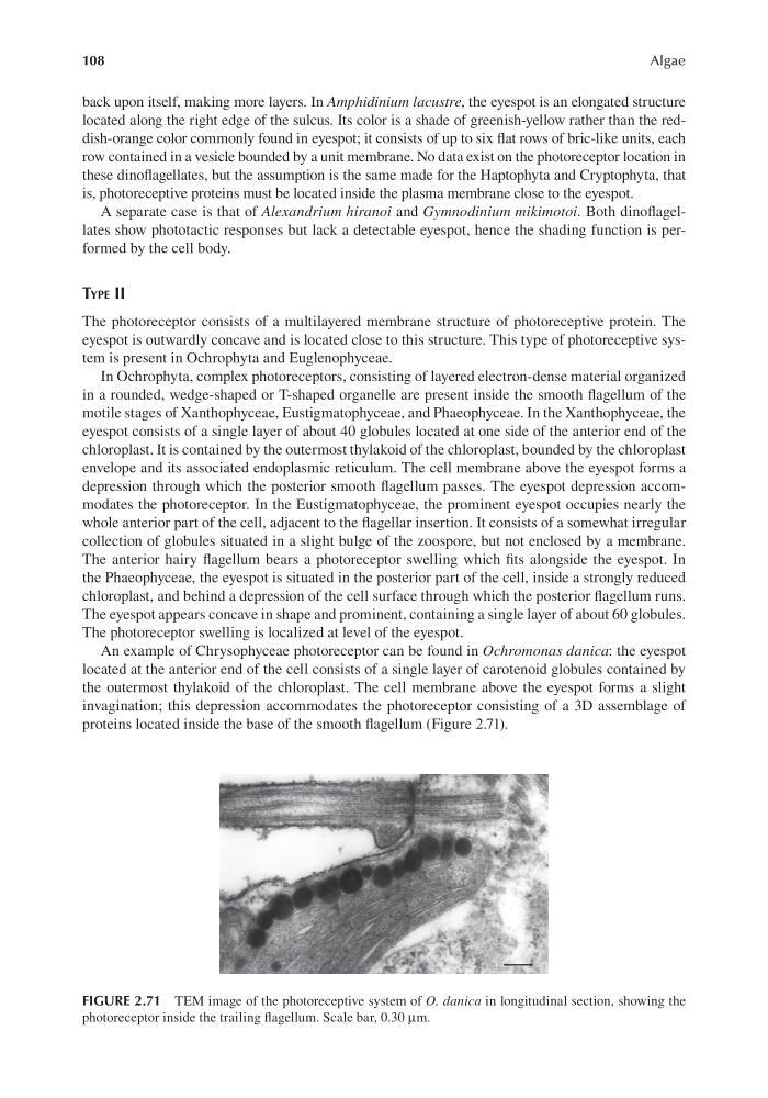
An example of Chrysophyceae photoreceptor can be found in Ochromonas danica: the eyespot
located at the anterior end of the cell consists of a single layer of carotenoid globules contained by
the outermost thylakoid of the chloroplast. The cell membrane above the eyespot forms a slight
invagination; this depression accommodates the photoreceptor consisting of a 3D assemblage of
proteins located inside the base of the smooth flagellum (Figure 2.71).
FIGURE 2.71 TEM image of the photoreceptive system of O. danica in longitudinal section, showing the
photoreceptor inside the trailing flagellum. Scale bar, 0.30 μm.
Algae
back upon itself, making more layers. In Amphidinium lacustre, the eyespot is an elongated structure
located along the right edge of the sulcus. Its color is a shade of greenish-yellow rather than the reddish-orange color commonly found in eyespot; it consists of up to six flat rows of bric-like units, each
row contained in a vesicle bounded by a unit membrane. No data exist on the photoreceptor location in
these dinoflagellates, but the assumption is the same made for the Haptophyta and Cryptophyta, that
is, photoreceptive proteins must be located inside the plasma membrane close to the eyespot.
A separate case is that of Alexandrium hiranoi and Gymnodinium mikimotoi. Both dinoflagellates show phototactic responses but lack a detectable eyespot, hence the shading function is performed by the cell body.
Type ii
The photoreceptor consists of a multilayered membrane structure of photoreceptive protein. The
eyespot is outwardly concave and is located close to this structure. This type of photoreceptive system is present in Ochrophyta and Euglenophyceae.
In Ochrophyta, complex photoreceptors, consisting of layered electron-dense material organized
in a rounded, wedge-shaped or T-shaped organelle are present inside the smooth flagellum of the
motile stages of Xanthophyceae, Eustigmatophyceae, and Phaeophyceae. In the Xanthophyceae, the
eyespot consists of a single layer of about 40 globules located at one side of the anterior end of the
chloroplast. It is contained by the outermost thylakoid of the chloroplast, bounded by the chloroplast
envelope and its associated endoplasmic reticulum. The cell membrane above the eyespot forms a
depression through which the posterior smooth flagellum passes. The eyespot depression accommodates the photoreceptor. In the Eustigmatophyceae, the prominent eyespot occupies nearly the
whole anterior part of the cell, adjacent to the flagellar insertion. It consists of a somewhat irregular
collection of globules situated in a slight bulge of the zoospore, but not enclosed by a membrane.
The anterior hairy flagellum bears a photoreceptor swelling which fits alongside the eyespot. In
the Phaeophyceae, the eyespot is situated in the posterior part of the cell, inside a strongly reduced
chloroplast, and behind a depression of the cell surface through which the posterior flagellum runs.
The eyespot appears concave in shape and prominent, containing a single layer of about 60 globules.
The photoreceptor swelling is localized at level of the eyespot.
An example of Chrysophyceae photoreceptor can be found in Ochromonas danica: the eyespot
located at the anterior end of the cell consists of a single layer of carotenoid globules contained by
the outermost thylakoid of the chloroplast. The cell membrane above the eyespot forms a slight
invagination; this depression accommodates the photoreceptor consisting of a 3D assemblage of
proteins located inside the base of the smooth flagellum (Figure 2.71).
FIGURE 2.71 TEM image of the photoreceptive system of O. danica in longitudinal section, showing the
photoreceptor inside the trailing flagellum. Scale bar, 0.30 μm.