

106
Algae
TYPES OF PHOTORECEPTIVE SYSTEMS
Depending on their organization and structure, photoreceptive systems can be grouped into three
main types.
Type i
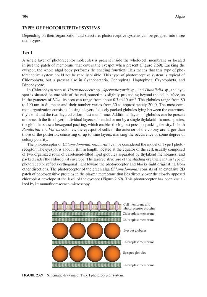
A single layer of photoreceptor molecules is present inside the whole-cell membrane or located
in just the patch of membrane that covers the eyespot when present (Figure 2.69). Lacking the
eyespot, the whole algal body performs the shading function. This means that this type of photoreceptive system could not be readily visible. This type of photoreceptive system is typical of
Chlorophyta, but is present also in Cyanobacteria, Ochrophyta, Haptophyta, Cryptophyta, and
Dinophyceae.
In Chlorophyta such as Haematococcus sp., Spermatozopsis sp., and Dunaliella sp., the eyespot is situated on one side of the cell, sometimes slightly protruding beyond the cell surface, as
in the gametes of Ulva; its area can range from about 0.3 to 10 μm 2 . The globules range from 80
to 190 nm in diameter and their number varies from 30 to approximately 2000. The most common organization consists of a single layer of closely packed globules lying between the outermost
thylakoid and the two-layered chloroplast membrane. Additional layers of globules can be present
underneath the first layer, individual layers subtended or not by a single thylakoid. In most species,
the globules show a hexagonal packing, which enables the highest possible packing density. In both
Pandorina and Volvox colonies, the eyespot of cells in the anterior of the colony are larger than
those of the posterior, consisting of up to nine layers, marking the occurrence of some degree of
colony polarity.
The photoreceptor of Chlamydomonas reinhardtii can be considered the model of Type I photoreceptor. The eyespot is about 1 μm in length, located at the equator of the cell, usually composed
of two organized rows of carotenoid-filled lipid globules separated by thylakoid membranes, and
packed under the chloroplast envelope. The layered structure of the shading organelle in this type of
photoreceptor reflects orthogonal light toward the photoreceptor and blocks light originating from
other directions. The photoreceptor of the green alga Chlamydomonas consists of an extensive 2D
patch of photosensitive proteins in the plasma membrane that lies directly over the closely apposed
chloroplast envelope at the level of the eyespot (Figure 2.69). This photoreceptor has been visualized by immunofluorescence microscopy.
Cell membrane and
photoreceptor proteins
Chloroplast membrane
Chloroplast membrane
Chloroplast membrane
Chloroplast membrane
Eyespot globules
Eyespot globules
FIGURE 2.69 Schematic drawing of Type I photoreceptor system.
Algae
TYPES OF PHOTORECEPTIVE SYSTEMS
Depending on their organization and structure, photoreceptive systems can be grouped into three
main types.
Type i
A single layer of photoreceptor molecules is present inside the whole-cell membrane or located
in just the patch of membrane that covers the eyespot when present (Figure 2.69). Lacking the
eyespot, the whole algal body performs the shading function. This means that this type of photoreceptive system could not be readily visible. This type of photoreceptive system is typical of
Chlorophyta, but is present also in Cyanobacteria, Ochrophyta, Haptophyta, Cryptophyta, and
Dinophyceae.
In Chlorophyta such as Haematococcus sp., Spermatozopsis sp., and Dunaliella sp., the eyespot is situated on one side of the cell, sometimes slightly protruding beyond the cell surface, as
in the gametes of Ulva; its area can range from about 0.3 to 10 μm 2 . The globules range from 80
to 190 nm in diameter and their number varies from 30 to approximately 2000. The most common organization consists of a single layer of closely packed globules lying between the outermost
thylakoid and the two-layered chloroplast membrane. Additional layers of globules can be present
underneath the first layer, individual layers subtended or not by a single thylakoid. In most species,
the globules show a hexagonal packing, which enables the highest possible packing density. In both
Pandorina and Volvox colonies, the eyespot of cells in the anterior of the colony are larger than
those of the posterior, consisting of up to nine layers, marking the occurrence of some degree of
colony polarity.
The photoreceptor of Chlamydomonas reinhardtii can be considered the model of Type I photoreceptor. The eyespot is about 1 μm in length, located at the equator of the cell, usually composed
of two organized rows of carotenoid-filled lipid globules separated by thylakoid membranes, and
packed under the chloroplast envelope. The layered structure of the shading organelle in this type of
photoreceptor reflects orthogonal light toward the photoreceptor and blocks light originating from
other directions. The photoreceptor of the green alga Chlamydomonas consists of an extensive 2D
patch of photosensitive proteins in the plasma membrane that lies directly over the closely apposed
chloroplast envelope at the level of the eyespot (Figure 2.69). This photoreceptor has been visualized by immunofluorescence microscopy.
Cell membrane and
photoreceptor proteins
Chloroplast membrane
Chloroplast membrane
Chloroplast membrane
Chloroplast membrane
Eyespot globules
Eyespot globules
FIGURE 2.69 Schematic drawing of Type I photoreceptor system.