

humans. The biological cell membrane is the first contact surface to the fluorinated
organic compounds. The interaction of PFOS and PFOA with DMPC bilayers
deposited on the gold electrode surface was investigated by means of in situ PM
IRRAS [40]. Water-soluble PFOS and PFOA molecules accumulate in the DMPC
monolayer at the air|water interface. LB-LS transfer was used to prepare DMPC
bilayers containing perdeuterated d 54 -DMPC in the inner leaflet and DMPCperfluorinated organic compound in the outer leaflet. The analysis of the PM
IRRA spectra in the CD stretching modes region provides information on the
conformation and tilt of the hydrocarbon chains in the lipid bilayer before and
after its interaction with perfluorinated organic compounds. Compared to a pure
DMPC bilayer, a small hypsochromic shift of the methylene stretching modes in the
membrane with incorporated PFOS (PFOA) is observed [40]. In this case the
intensities of the methylene stretching modes are lower than in the pure DMPC
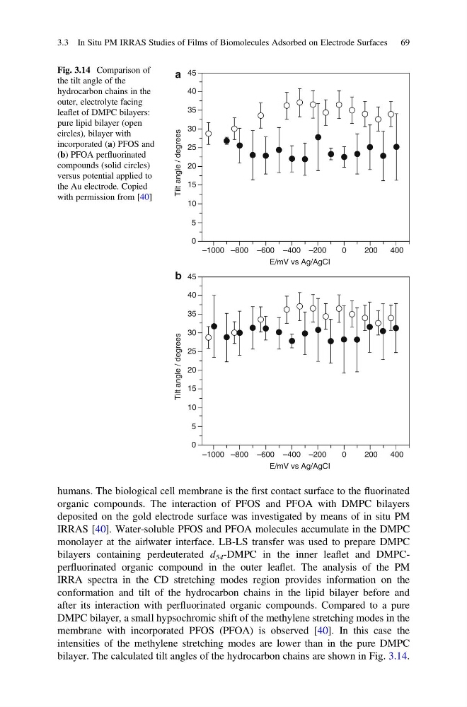
bilayer. The calculated tilt angles of the hydrocarbon chains are shown in Fig. 3.14.
400
200
0
–200
–400
E/mV vs Ag/AgCI
–600
–800
0
5
10
15
Tilt angle / degrees
20
25
30
35
40
45
–1000
400
200
0
–200
–400
E/mV vs Ag/AgCI
–600
–800
0
5
10
15
Tilt angle / degrees
20
25
30
35
40
45
–1000
a
b
Fig. 3.14 Comparison of
the tilt angle of the
hydrocarbon chains in the
outer, electrolyte facing
leaflet of DMPC bilayers:
pure lipid bilayer (open
circles), bilayer with
incorporated (a) PFOS and
(b) PFOA perfluorinated
compounds (solid circles)
versus potential applied to
the Au electrode. Copied
with permission from [40]
3.3 In Situ PM IRRAS Studies of Films of Biomolecules Adsorbed on Electrode Surfaces 69
organic compounds. The interaction of PFOS and PFOA with DMPC bilayers
deposited on the gold electrode surface was investigated by means of in situ PM
IRRAS [40]. Water-soluble PFOS and PFOA molecules accumulate in the DMPC
monolayer at the air|water interface. LB-LS transfer was used to prepare DMPC
bilayers containing perdeuterated d 54 -DMPC in the inner leaflet and DMPCperfluorinated organic compound in the outer leaflet. The analysis of the PM
IRRA spectra in the CD stretching modes region provides information on the
conformation and tilt of the hydrocarbon chains in the lipid bilayer before and
after its interaction with perfluorinated organic compounds. Compared to a pure
DMPC bilayer, a small hypsochromic shift of the methylene stretching modes in the
membrane with incorporated PFOS (PFOA) is observed [40]. In this case the
intensities of the methylene stretching modes are lower than in the pure DMPC
bilayer. The calculated tilt angles of the hydrocarbon chains are shown in Fig. 3.14.
400
200
0
–200
–400
E/mV vs Ag/AgCI
–600
–800
0
5
10
15
Tilt angle / degrees
20
25
30
35
40
45
–1000
400
200
0
–200
–400
E/mV vs Ag/AgCI
–600
–800
0
5
10
15
Tilt angle / degrees
20
25
30
35
40
45
–1000
a
b
Fig. 3.14 Comparison of
the tilt angle of the
hydrocarbon chains in the
outer, electrolyte facing
leaflet of DMPC bilayers:
pure lipid bilayer (open
circles), bilayer with
incorporated (a) PFOS and
(b) PFOA perfluorinated
compounds (solid circles)
versus potential applied to
the Au electrode. Copied
with permission from [40]
3.3 In Situ PM IRRAS Studies of Films of Biomolecules Adsorbed on Electrode Surfaces 69