

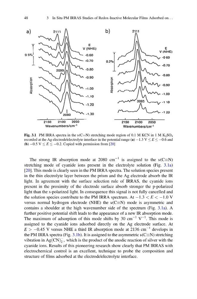
The strong IR absorption mode at 2080 cm
À1 is assigned to the ν(CN)
stretching mode of cyanide ions present in the electrolyte solution (Fig. 3.1a)
[20]. This mode is clearly seen in the PM IRRA spectra. The solution species present
in the thin electrolyte layer between the prism and the Ag electrode absorb the IR
light. In agreement with the surface selection rule of IRRAS, the cyanide ions
present in the proximity of the electrode surface absorb stronger the p-polarized
light than the s-polarized light. In consequence this signal is not fully cancelled and
the solution species contribute to the PM IRRA spectrum. At À1.3 < E < À1.0 V
versus normal hydrogen electrode (NHE) the ν(CN) mode is asymmetric and
contains a shoulder at the high wavenumber side of the spectrum (Fig. 3.1a). A
further positive potential shift leads to the appearance of a new IR absorption mode.
The maximum of adsorption of this mode shifts by 30 cm
À1 V
À1 . This mode is
assigned to the cyanide ions adsorbed directly on the Ag electrode surface. At
E > À0.45 V versus NHE a third IR absorption mode at 2136 cm
À1 develops in
the PM IRRA spectra (Fig. 3.1b). It is assigned to the asymmetric ν(CN) stretching
vibration in Ag CN
ð Þ
À
2 , which is the product of the anodic reaction of silver with the
cyanide ions. Results of this pioneering research show clearly that PM IRRAS with
electrochemical control is an excellent, technique to probe the composition and
structure of films adsorbed at the electrode|electrolyte interface.
Fig. 3.1 PM IRRA spectra in the ν(CN) stretching mode region of 0.1 M KCN in 1 M K 2 SO 4
recorded at the Ag electrode|electrolyte interface in the potential range (a) À1.3 V E À0.6 and
(b) À0.5 V E À0.2. Copied with permission from [20]
48
3 In Situ PM IRRAS Studies of Redox-Inactive Molecular Films Adsorbed on. . .
À1 is assigned to the ν(CN)
stretching mode of cyanide ions present in the electrolyte solution (Fig. 3.1a)
[20]. This mode is clearly seen in the PM IRRA spectra. The solution species present
in the thin electrolyte layer between the prism and the Ag electrode absorb the IR
light. In agreement with the surface selection rule of IRRAS, the cyanide ions
present in the proximity of the electrode surface absorb stronger the p-polarized
light than the s-polarized light. In consequence this signal is not fully cancelled and
the solution species contribute to the PM IRRA spectrum. At À1.3 < E < À1.0 V
versus normal hydrogen electrode (NHE) the ν(CN) mode is asymmetric and
contains a shoulder at the high wavenumber side of the spectrum (Fig. 3.1a). A
further positive potential shift leads to the appearance of a new IR absorption mode.
The maximum of adsorption of this mode shifts by 30 cm
À1 V
À1 . This mode is
assigned to the cyanide ions adsorbed directly on the Ag electrode surface. At
E > À0.45 V versus NHE a third IR absorption mode at 2136 cm
À1 develops in
the PM IRRA spectra (Fig. 3.1b). It is assigned to the asymmetric ν(CN) stretching
vibration in Ag CN
ð Þ
À
2 , which is the product of the anodic reaction of silver with the
cyanide ions. Results of this pioneering research show clearly that PM IRRAS with
electrochemical control is an excellent, technique to probe the composition and
structure of films adsorbed at the electrode|electrolyte interface.
Fig. 3.1 PM IRRA spectra in the ν(CN) stretching mode region of 0.1 M KCN in 1 M K 2 SO 4
recorded at the Ag electrode|electrolyte interface in the potential range (a) À1.3 V E À0.6 and
(b) À0.5 V E À0.2. Copied with permission from [20]
48
3 In Situ PM IRRAS Studies of Redox-Inactive Molecular Films Adsorbed on. . .