

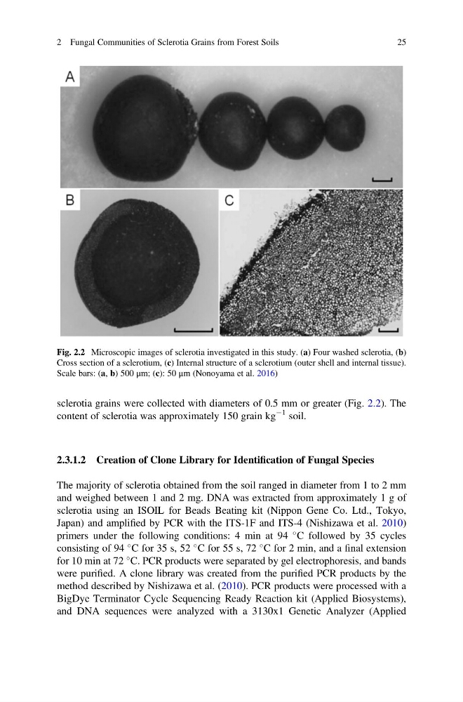
sclerotia grains were collected with diameters of 0.5 mm or greater (Fig. 2.2). The
content of sclerotia was approximately 150 grain kg
À1 soil.
2.3.1.2 Creation of Clone Library for Identification of Fungal Species
The majority of sclerotia obtained from the soil ranged in diameter from 1 to 2 mm
and weighed between 1 and 2 mg. DNA was extracted from approximately 1 g of
sclerotia using an ISOIL for Beads Beating kit (Nippon Gene Co. Ltd., Tokyo,
Japan) and amplified by PCR with the ITS-1F and ITS-4 (Nishizawa et al. 2010)
primers under the following conditions: 4 min at 94
C followed by 35 cycles
consisting of 94
C for 35 s, 52
C for 55 s, 72
C for 2 min, and a final extension
for 10 min at 72
C. PCR products were separated by gel electrophoresis, and bands
were purified. A clone library was created from the purified PCR products by the
method described by Nishizawa et al. (2010). PCR products were processed with a
BigDye Terminator Cycle Sequencing Ready Reaction kit (Applied Biosystems),
and DNA sequences were analyzed with a 3130x1 Genetic Analyzer (Applied
Fig. 2.2 Microscopic images of sclerotia investigated in this study. (a) Four washed sclerotia, (b)
Cross section of a sclerotium, (c) Internal structure of a sclerotium (outer shell and internal tissue).
Scale bars: (a, b) 500 μm; (c): 50 μm (Nonoyama et al. 2016)
2 Fungal Communities of Sclerotia Grains from Forest Soils
25
content of sclerotia was approximately 150 grain kg
À1 soil.
2.3.1.2 Creation of Clone Library for Identification of Fungal Species
The majority of sclerotia obtained from the soil ranged in diameter from 1 to 2 mm
and weighed between 1 and 2 mg. DNA was extracted from approximately 1 g of
sclerotia using an ISOIL for Beads Beating kit (Nippon Gene Co. Ltd., Tokyo,
Japan) and amplified by PCR with the ITS-1F and ITS-4 (Nishizawa et al. 2010)
primers under the following conditions: 4 min at 94
C followed by 35 cycles
consisting of 94
C for 35 s, 52
C for 55 s, 72
C for 2 min, and a final extension
for 10 min at 72
C. PCR products were separated by gel electrophoresis, and bands
were purified. A clone library was created from the purified PCR products by the
method described by Nishizawa et al. (2010). PCR products were processed with a
BigDye Terminator Cycle Sequencing Ready Reaction kit (Applied Biosystems),
and DNA sequences were analyzed with a 3130x1 Genetic Analyzer (Applied
Fig. 2.2 Microscopic images of sclerotia investigated in this study. (a) Four washed sclerotia, (b)
Cross section of a sclerotium, (c) Internal structure of a sclerotium (outer shell and internal tissue).
Scale bars: (a, b) 500 μm; (c): 50 μm (Nonoyama et al. 2016)
2 Fungal Communities of Sclerotia Grains from Forest Soils
25