

on the displayed monolith specimens of forest soils in museums, characterized by the
black spherical grain; and on specimens of thin sections, characterized by their
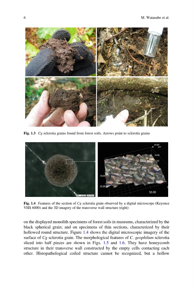
hollowed round structure. Figure 1.4 shows the digital microscopic imagery of the
surface of Cg sclerotia grain. The morphological features of C. geophilum sclerotia
sliced into half pieces are shown in Figs. 1.5 and 1.6. They have honeycomb
structure in their transverse wall constructed by the empty cells contacting each
other. Histopathological coiled structure cannot be recognized, but a hollow
Fig. 1.3 Cg sclerotia grains found from forest soils. Arrows point to sclerotia grains
Fig. 1.4 Features of the section of Cg sclerotia grain observed by a digital microscope (Keyence
VHS 6000) and the 3D imagery of the transverse wall structure (right)
6
M. Watanabe et al.
black spherical grain; and on specimens of thin sections, characterized by their
hollowed round structure. Figure 1.4 shows the digital microscopic imagery of the
surface of Cg sclerotia grain. The morphological features of C. geophilum sclerotia
sliced into half pieces are shown in Figs. 1.5 and 1.6. They have honeycomb
structure in their transverse wall constructed by the empty cells contacting each
other. Histopathological coiled structure cannot be recognized, but a hollow
Fig. 1.3 Cg sclerotia grains found from forest soils. Arrows point to sclerotia grains
Fig. 1.4 Features of the section of Cg sclerotia grain observed by a digital microscope (Keyence
VHS 6000) and the 3D imagery of the transverse wall structure (right)
6
M. Watanabe et al.