

34
Electromagnetic Fields in Biological Systems
dB
0 = 1.25 W/kg
x
−6
−12
−18
−24
z
−30
y
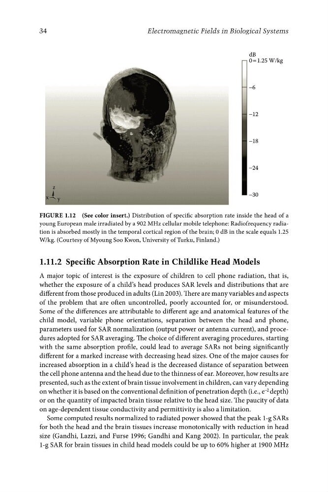
FigurE 1.12 (See color insert.) Distribution of specific absorption rate inside the head of a
young European male irradiated by a 902 MHz cellular mobile telephone: Radiofrequency radiation is absorbed mostly in the temporal cortical region of the brain; 0 dB in the scale equals 1.25
W/kg. (Courtesy of Myoung Soo Kwon, University of Turku, Finland.)
1.11.2 Specific Absorption Rate in Childlike Head Models
A major topic of interest is the exposure of children to cell phone radiation, that is,
whether the exposure of a child’s head produces SAR levels and distributions that are
different from those produced in adults (Lin 2003). There are many variables and aspects
of the problem that are often uncontrolled, poorly accounted for, or misunderstood.
Some of the differences are attributable to different age and anatomical features of the
child model, variable phone orientations, separation between the head and phone,
parameters used for SAR normalization (output power or antenna current), and procedures adopted for SAR averaging. The choice of different averaging procedures, starting
with the same absorption profile, could lead to average SARs not being significantly
different for a marked increase with decreasing head sizes. One of the major causes for
increased absorption in a child’s head is the decreased distance of separation between
the cell phone antenna and the head due to the thinness of ear. Moreover, how results are
presented, such as the extent of brain tissue involvement in children, can vary depending
on whether it is based on the conventional definition of penetration depth (i.e., e −2 depth)
or on the quantity of impacted brain tissue relative to the head size. The paucity of data
on age-dependent tissue conductivity and permittivity is also a limitation.
Some computed results normalized to radiated power showed that the peak 1-g SARs
for both the head and the brain tissues increase monotonically with reduction in head
size (Gandhi, Lazzi, and Furse 1996; Gandhi and Kang 2002). In particular, the peak
1-g SAR for brain tissues in child head models could be up to 60% higher at 1900 MHz
Electromagnetic Fields in Biological Systems
dB
0 = 1.25 W/kg
x
−6
−12
−18
−24
z
−30
y
FigurE 1.12 (See color insert.) Distribution of specific absorption rate inside the head of a
young European male irradiated by a 902 MHz cellular mobile telephone: Radiofrequency radiation is absorbed mostly in the temporal cortical region of the brain; 0 dB in the scale equals 1.25
W/kg. (Courtesy of Myoung Soo Kwon, University of Turku, Finland.)
1.11.2 Specific Absorption Rate in Childlike Head Models
A major topic of interest is the exposure of children to cell phone radiation, that is,
whether the exposure of a child’s head produces SAR levels and distributions that are
different from those produced in adults (Lin 2003). There are many variables and aspects
of the problem that are often uncontrolled, poorly accounted for, or misunderstood.
Some of the differences are attributable to different age and anatomical features of the
child model, variable phone orientations, separation between the head and phone,
parameters used for SAR normalization (output power or antenna current), and procedures adopted for SAR averaging. The choice of different averaging procedures, starting
with the same absorption profile, could lead to average SARs not being significantly
different for a marked increase with decreasing head sizes. One of the major causes for
increased absorption in a child’s head is the decreased distance of separation between
the cell phone antenna and the head due to the thinness of ear. Moreover, how results are
presented, such as the extent of brain tissue involvement in children, can vary depending
on whether it is based on the conventional definition of penetration depth (i.e., e −2 depth)
or on the quantity of impacted brain tissue relative to the head size. The paucity of data
on age-dependent tissue conductivity and permittivity is also a limitation.
Some computed results normalized to radiated power showed that the peak 1-g SARs
for both the head and the brain tissues increase monotonically with reduction in head
size (Gandhi, Lazzi, and Furse 1996; Gandhi and Kang 2002). In particular, the peak
1-g SAR for brain tissues in child head models could be up to 60% higher at 1900 MHz