

15
1
x-axis
50
0.9
y-axis
0.8
100
100
200
300
400
500
150
10
0.7
0.6
0.5
0.4
0.6
400
0.2
450
0.1
500
0
0 −8 −6 −4 −2
0
2
4
6
8
Location (cm)
15
1
x-axis
50
0.9
y-axis
100
0.8
SAR/SAR max
200
250
300
5
350
100
150
SAR/SAR max
0.7
0.6
0.5
0.4
0.6
400
0.2
450
0.1
500
0
0
200
300
400
500
−8 −6 −4 −2
0
2
4
6
8
Location (cm)
15
1
x-axis
50
0.9
y-axis
100
0.8
10
200
1
250
300
5
350
100
200
300
400
500
SAR/SAR max
0.7
0.6
0.5
0.4
0.6
400
0.2
0.1
450
500
0
0 −8 −6 −4 −2
0
2
4
6
8
150
10
200
250
300
5
350
Location (cm)
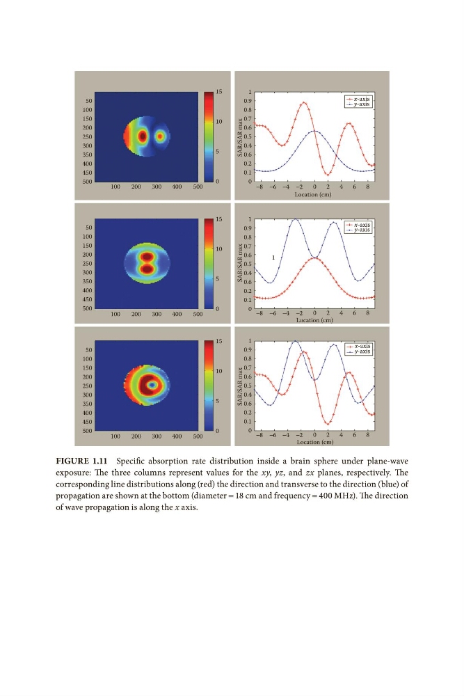
Figure 1.11 Specific absorption rate distribution inside a brain sphere under plane-wave
exposure: The three columns represent values for the xy, yz, and zx planes, respectively. The
corresponding line distributions along (red) the direction and transverse to the direction (blue) of
propagation are shown at the bottom (diameter = 18 cm and frequency = 400 MHz). The direction
of wave propagation is along the x axis.
1
x-axis
50
0.9
y-axis
0.8
100
100
200
300
400
500
150
10
0.7
0.6
0.5
0.4
0.6
400
0.2
450
0.1
500
0
0 −8 −6 −4 −2
0
2
4
6
8
Location (cm)
15
1
x-axis
50
0.9
y-axis
100
0.8
SAR/SAR max
200
250
300
5
350
100
150
SAR/SAR max
0.7
0.6
0.5
0.4
0.6
400
0.2
450
0.1
500
0
0
200
300
400
500
−8 −6 −4 −2
0
2
4
6
8
Location (cm)
15
1
x-axis
50
0.9
y-axis
100
0.8
10
200
1
250
300
5
350
100
200
300
400
500
SAR/SAR max
0.7
0.6
0.5
0.4
0.6
400
0.2
0.1
450
500
0
0 −8 −6 −4 −2
0
2
4
6
8
150
10
200
250
300
5
350
Location (cm)
Figure 1.11 Specific absorption rate distribution inside a brain sphere under plane-wave
exposure: The three columns represent values for the xy, yz, and zx planes, respectively. The
corresponding line distributions along (red) the direction and transverse to the direction (blue) of
propagation are shown at the bottom (diameter = 18 cm and frequency = 400 MHz). The direction
of wave propagation is along the x axis.