

352
Electromagnetic Fields in Biological Systems
0.000739
0.00191
0.00335
0.0051
0.00724
0.00985
0.013
0.0169
0.0217
0.0275
0.0346
0.0914
W/kg
0
Figure 6.6 SAR distribution within the sagittal plane of a pregnant woman SILVY and the
fetus during 4-T magnetic resonance imaging (MRI) with 170 MHz radio-frequency electromagnetic field (from PF EMF). (Data from Pediaditis, M., N. Leitgeb, and R. Cech. 2008. Phys Med
Biol 53:7187–95.)
B/dt
10
100
1000
T/s
10000
1 st LCOM
t s, eff
C S t h r e s h o l d
P N S t h r e s h o l d
NOM
2 nd LCOM
0.01
0.1
1
10 ms
1
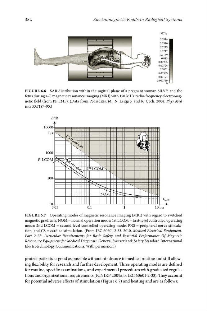
Figure 6.7 Operating modes of magnetic resonance imaging (MRI) with regard to switched
magnetic gradients. NOM = normal operation mode; 1st LCOM = first-level controlled operating
mode; 2nd LCOM = second-level controlled operating mode; PNS = peripheral nerve stimulation; and CS = cardiac stimulation. (From IEC 60601-2-33. 2010. Medical Electrical Equipment.
Part 2–33: Particular Requirements for Basic Safety and Essential Performance Of Magnetic
Resonance Equipment for Medical Diagnosis. Geneva, Switzerland: Safety Standard International
Electrotechnology Communications. With permission.)
protect patients as good as possible without hindrance to medical routine and still allowing flexibility for research and further development. Three operating modes are defined
for routine, specific examinations, and experimental procedures with graduated regulations and organizational requirements (ICNIRP 2009a,b; IEC 60601-2-33). They account
for potential adverse effects of stimulation (Figure 6.7) and heating and are as follows:
Electromagnetic Fields in Biological Systems
0.000739
0.00191
0.00335
0.0051
0.00724
0.00985
0.013
0.0169
0.0217
0.0275
0.0346
0.0914
W/kg
0
Figure 6.6 SAR distribution within the sagittal plane of a pregnant woman SILVY and the
fetus during 4-T magnetic resonance imaging (MRI) with 170 MHz radio-frequency electromagnetic field (from PF EMF). (Data from Pediaditis, M., N. Leitgeb, and R. Cech. 2008. Phys Med
Biol 53:7187–95.)
B/dt
10
100
1000
T/s
10000
1 st LCOM
t s, eff
C S t h r e s h o l d
P N S t h r e s h o l d
NOM
2 nd LCOM
0.01
0.1
1
10 ms
1
Figure 6.7 Operating modes of magnetic resonance imaging (MRI) with regard to switched
magnetic gradients. NOM = normal operation mode; 1st LCOM = first-level controlled operating
mode; 2nd LCOM = second-level controlled operating mode; PNS = peripheral nerve stimulation; and CS = cardiac stimulation. (From IEC 60601-2-33. 2010. Medical Electrical Equipment.
Part 2–33: Particular Requirements for Basic Safety and Essential Performance Of Magnetic
Resonance Equipment for Medical Diagnosis. Geneva, Switzerland: Safety Standard International
Electrotechnology Communications. With permission.)
protect patients as good as possible without hindrance to medical routine and still allowing flexibility for research and further development. Three operating modes are defined
for routine, specific examinations, and experimental procedures with graduated regulations and organizational requirements (ICNIRP 2009a,b; IEC 60601-2-33). They account
for potential adverse effects of stimulation (Figure 6.7) and heating and are as follows: