

Mobile Communication Fields in Biological Systems
271
(a)
(b)
(c)
(d)
(e)
(f)
(g)
Table 5.1 Female Analytical Models Used in the Literature
Age
Height
Weight
Voxel Size
Number of
Reference
(years)
(cm)
(kg)
(mm 3 )
Tissue Types
Mazzurana et al. (2004)
30
163
47
2 × 2 × 2
n/a
Liu, Collins, and Smith (2005)
n/a
165
85.5
5 × 5 × 5
36
Dimbylow (2005a,b)
23
165
58
2 × 2 × 2
41
Nagaoka et al. (2004)
22
160
53
2 × 2 × 2
51
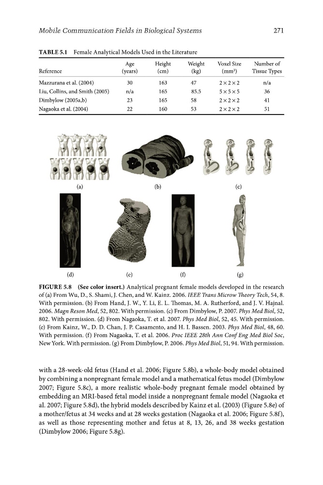
Figure 5.8 (See color insert.) Analytical pregnant female models developed in the research
of (a) From Wu, D., S. Shami, J. Chen, and W. Kainz. 2006. IEEE Trans Microw Theory Tech, 54, 8.
With permission. (b) From Hand, J. W., Y. Li, E. L. Thomas, M. A. Rutherford, and J. V. Hajnal.
2006. Magn Reson Med, 52, 802. With permission. (c) From Dimbylow, P. 2007. Phys Med Biol, 52,
802. With permission. (d) From Nagaoka, T. et al. 2007. Phys Med Biol, 52, 45. With permission.
(e) From Kainz, W., D. D. Chan, J. P. Casamento, and H. I. Bassen. 2003. Phys Med Biol, 48, 60.
With permission. (f) From Nagaoka, T. et al. 2006. Proc IEEE 28th Ann Conf Eng Med Biol Soc,
New York. With permission. (g) From Dimbylow, P. 2006. Phys Med Biol, 51, 94. With permission.
with a 28-week-old fetus (Hand et al. 2006; Figure 5.8b), a whole-body model obtained
by combining a nonpregnant female model and a mathematical fetus model (Dimbylow
2007; Figure 5.8c), a more realistic whole-body pregnant female model obtained by
embedding an MRI-based fetal model inside a nonpregnant female model (Nagaoka et
al. 2007; Figure 5.8d), the hybrid models described by Kainz et al. (2003) (Figure 5.8e) of
a mother/fetus at 34 weeks and at 28 weeks gestation (Nagaoka et al. 2006; Figure 5.8f),
as well as those representing mother and fetus at 8, 13, 26, and 38 weeks gestation
(Dimbylow 2006; Figure 5.8g).
271
(a)
(b)
(c)
(d)
(e)
(f)
(g)
Table 5.1 Female Analytical Models Used in the Literature
Age
Height
Weight
Voxel Size
Number of
Reference
(years)
(cm)
(kg)
(mm 3 )
Tissue Types
Mazzurana et al. (2004)
30
163
47
2 × 2 × 2
n/a
Liu, Collins, and Smith (2005)
n/a
165
85.5
5 × 5 × 5
36
Dimbylow (2005a,b)
23
165
58
2 × 2 × 2
41
Nagaoka et al. (2004)
22
160
53
2 × 2 × 2
51
Figure 5.8 (See color insert.) Analytical pregnant female models developed in the research
of (a) From Wu, D., S. Shami, J. Chen, and W. Kainz. 2006. IEEE Trans Microw Theory Tech, 54, 8.
With permission. (b) From Hand, J. W., Y. Li, E. L. Thomas, M. A. Rutherford, and J. V. Hajnal.
2006. Magn Reson Med, 52, 802. With permission. (c) From Dimbylow, P. 2007. Phys Med Biol, 52,
802. With permission. (d) From Nagaoka, T. et al. 2007. Phys Med Biol, 52, 45. With permission.
(e) From Kainz, W., D. D. Chan, J. P. Casamento, and H. I. Bassen. 2003. Phys Med Biol, 48, 60.
With permission. (f) From Nagaoka, T. et al. 2006. Proc IEEE 28th Ann Conf Eng Med Biol Soc,
New York. With permission. (g) From Dimbylow, P. 2006. Phys Med Biol, 51, 94. With permission.
with a 28-week-old fetus (Hand et al. 2006; Figure 5.8b), a whole-body model obtained
by combining a nonpregnant female model and a mathematical fetus model (Dimbylow
2007; Figure 5.8c), a more realistic whole-body pregnant female model obtained by
embedding an MRI-based fetal model inside a nonpregnant female model (Nagaoka et
al. 2007; Figure 5.8d), the hybrid models described by Kainz et al. (2003) (Figure 5.8e) of
a mother/fetus at 34 weeks and at 28 weeks gestation (Nagaoka et al. 2006; Figure 5.8f),
as well as those representing mother and fetus at 8, 13, 26, and 38 weeks gestation
(Dimbylow 2006; Figure 5.8g).