

268
(a)
(b)
(c)
(d)
(e)
Electromagnetic Fields in Biological Systems
developed by the National Institute of Information and Communication Technology
(NICT) (Nagaoka et al. 2004; Figure 5.4d). The model consists of 2 mm edge voxels and
is segmented to 51 tissue types. Examples of body models in postures other than standing can be found in Allen et al. (2003, 2005), Findlay and Dimbylow (2005, 2006), and
Uusitupa et al. (2010). A review of voxel-based models developed for a range of applications can be found in Caon (2004).
Since the power absorbed in the human head is considered to be the most crucial
parameter in assessing the potential hazard caused by the use of mobile communication
devices, several research studies have focused on modeling the human head (Bernardi,
Cavagnaro, and Pisa 1996; Burkhardt and Kuster 2000; Christ and Kuster 2005; Groot
et al. 1997; Hirata, Matsuyama, and Shiozawa 2000; Hombach et al. 1996; Meier et al. 1997;
Okoniewski and Stuchly 1996; Schoenborn, Burkhardt, and Kuster 1998; Watanabe et al.
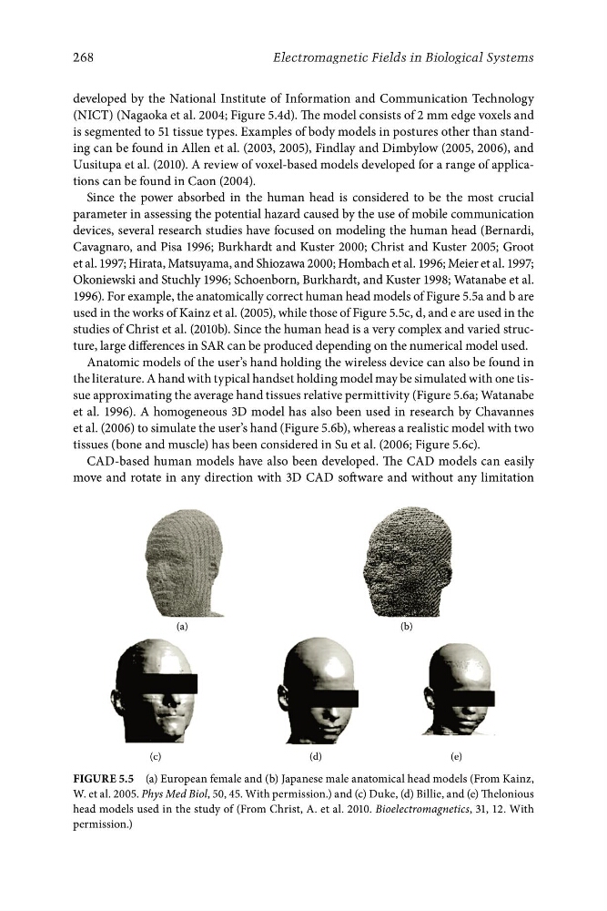
1996). For example, the anatomically correct human head models of Figure 5.5a and b are
used in the works of Kainz et al. (2005), while those of Figure 5.5c, d, and e are used in the
studies of Christ et al. (2010b). Since the human head is a very complex and varied structure, large differences in SAR can be produced depending on the numerical model used.
Anatomic models of the user’s hand holding the wireless device can also be found in
the literature. A hand with typical handset holding model may be simulated with one tissue approximating the average hand tissues relative permittivity (Figure 5.6a; Watanabe
et al. 1996). A homogeneous 3D model has also been used in research by Chavannes
et al. (2006) to simulate the user’s hand (Figure 5.6b), whereas a realistic model with two
tissues (bone and muscle) has been considered in Su et al. (2006; Figure 5.6c).
CAD-based human models have also been developed. The CAD models can easily
move and rotate in any direction with 3D CAD software and without any limitation
Figure 5.5 (a) European female and (b) Japanese male anatomical head models (From Kainz,
W. et al. 2005. Phys Med Biol, 50, 45. With permission.) and (c) Duke, (d) Billie, and (e) Thelonious
head models used in the study of (From Christ, A. et al. 2010. Bioelectromagnetics, 31, 12. With
permission.)