

227
Interaction of Extremely Low–Frequency Electromagnetic Fields
Brain
y
Heart
Lung
Liver
Front
Intestine
Front view
Side view
z
x
z
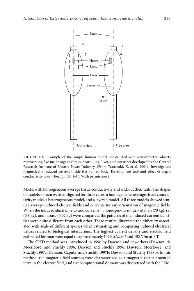
Figure 4.6 Example of the simple human model constructed with axisymmetric objects
representing five major organs (brain, heart, lung, liver, and intestine) developed by the Central
Research Institute of Electric Power Industry. (From Yamazaki, K. et al. 2001a. Investigation
magnetically induced current inside the human body: Development tool and effect of organ
conductivity. Electr Eng Jpn 134:1–10. With permission.)
MRIs, with homogeneous average tissue conductivity and without their tails. The shapes
of models of man were configured for three cases: a homogeneous average tissue conductivity model, a heterogeneous model, and a layered model. All three models showed similar average induced electric fields and currents for any orientation of magnetic fields.
When the induced electric fields and currents in homogenous models of man (70 kg), rat
(0.3 kg), and mouse (0.02 kg) were compared, the patterns of the induced current densities were quite different from each other. These results illustrated the difficulty associated with scale of different species when estimating and comparing induced electrical
values related to biological interactions. The highest current density and electric field
estimated for man were equal to approximately 1090 μA/cm 2 and 152 V/m at 1 T.
The SPFD method was introduced in 1996 by Dawson and coworkers (Dawson, de
Moerloose, and Stuchly 1996; Dawson and Stuchly 1996; Dawson, Moerloose, and
Stuchly 1997a; Dawson, Caputa, and Stuchly 1997b; Dawson and Stuchly 1998b). In this
method, the magnetic field sources were characterized as a magnetic vector potential
term in the electric field, and the computational domain was discretized with the FEM.