

209
Interaction of Extremely Low–Frequency Electromagnetic Fields
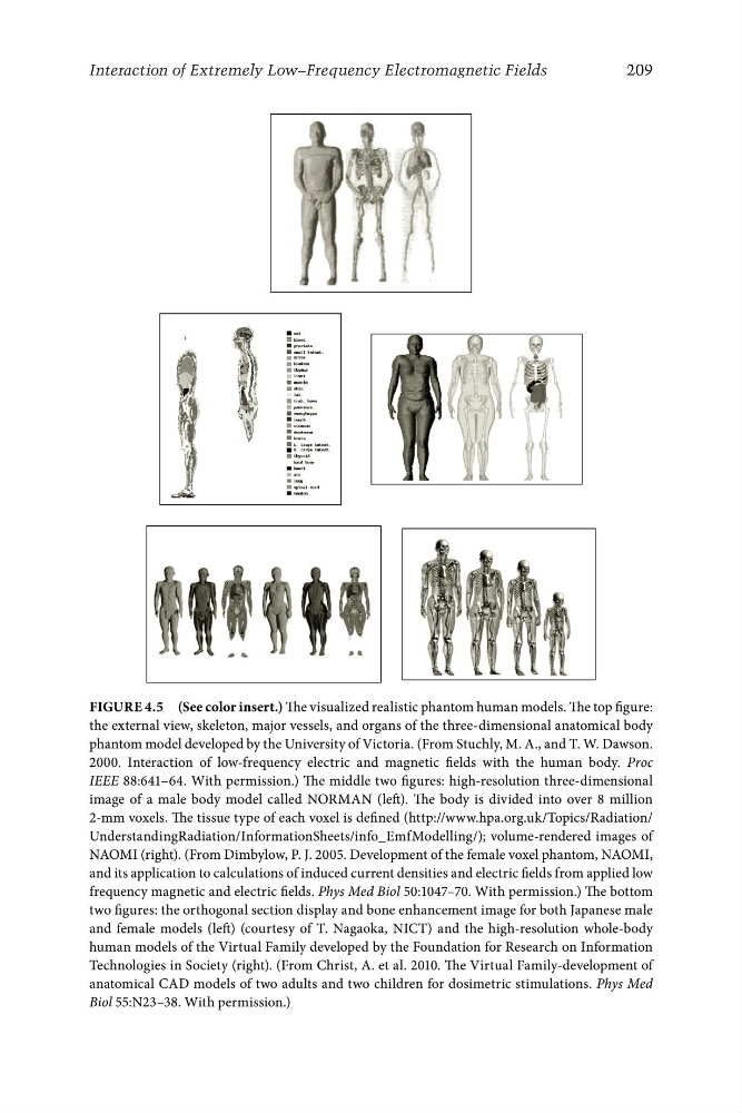
Figure 4.5 (See color insert.) The visualized realistic phantom human models. The top figure:
the external view, skeleton, major vessels, and organs of the three-dimensional anatomical body
phantom model developed by the University of Victoria. (From Stuchly, M. A., and T. W. Dawson.
2000. Interaction of low-frequency electric and magnetic fields with the human body. Proc
IEEE 88:641–64. With permission.) The middle two figures: high-resolution three-dimensional
image of a male body model called NORMAN (left). The body is divided into over 8 million
2-mm voxels. The tissue type of each voxel is defined (http://www.hpa.org.uk/Topics/Radiation/
UnderstandingRadiation/InformationSheets/info_EmfModelling/); volume-rendered images of
NAOMI (right). (From Dimbylow, P. J. 2005. Development of the female voxel phantom, NAOMI,
and its application to calculations of induced current densities and electric fields from applied low
frequency magnetic and electric fields. Phys Med Biol 50:1047–70. With permission.) The bottom
two figures: the orthogonal section display and bone enhancement image for both Japanese male
and female models (left) (courtesy of T. Nagaoka, NICT) and the high-resolution whole-body
human models of the Virtual Family developed by the Foundation for Research on Information
Technologies in Society (right). (From Christ, A. et al. 2010. The Virtual Family-development of
anatomical CAD models of two adults and two children for dosimetric stimulations. Phys Med
Biol 55:N23–38. With permission.)