

161
Static, Low-Frequency, and Pulsed Magnetic Fields
the peak ipsilesional sensorimotor cortex activity shifted posteriorly in more impaired
patients. Critical new findings were that concurrent TMS-fMRI results correlated with
the level of both clinical impairment and neurophysiological impairment. Specifically,
greater clinical and neurophysiological impairment was associated with a stronger facilitating influence of cPMd TMS on blood oxygenation level–dependent (BOLD) signal in
posterior parts of ipsilesional sensorimotor cortex during hand grip, corresponding to
the posteriorly shifted sensorimotor activity seen in more impaired patients. The cPMd
TMS was not found to influence activity in other brain regions in either hemisphere.
The authors suggested that this state-dependent influence on ipsilesional sensorimotor
regions provides a mechanism by which cPMd supports recovered function after stroke.
Dimyan and Cohen (2010) discussed the use of TMS in the diagnosis, prognosis,
and therapy of poststroke motor disability and emphasized that TMS is a useful tool
to explore mechanisms of neuroplasticity during spontaneous and treatment-induced
recovery of motor function to develop more rational and clinically useful interventions
for stroke rehabilitation.
The spatial extent of the effects of TMS on neural tissue is only coarsely understood. One key problem is the realistic calculation of the electric field induced in the
brain, which proves difficult due to the complex gyral folding pattern that results in
an inhomogeneous conductivity distribution within the skull. Thielscher, Opitz, and
Windhoff (2011) estimated the electric field induced in the brain using the finite element method (FEM) together with a high-resolution volume mesh of the human head
to better characterize the field induced in cortical gray matter (GM). The volume mesh
was constructed from T1-weighted structural MRI to ensure an anatomically accurate
modeling of the gyrification pattern. Five tissue types were taken into account, corresponding to skin, skull, cerebrospinal fluid (CSF) including the ventricles, cortical
GM, and cortical white matter. The authors characterized the effect of current direction on the electric field distribution in GM. The field strength in GM was increased
by up to 51% when the induced currents were perpendicular to the local gyrus orientation. This effect was mainly restricted to gyral crowns and lips, and did not extend
into the sulcal walls. As a result, the focality of the fields induced in GM was increased.
The authors speculated that this enhancement effect might in part explain the dependency of stimulation thresholds on coil orientation, which is commonly observed in
TMS motor cortex studies. In contrast to the clear-cut effects of the gyrification pattern on induced field strength, current directions were predominantly influenced by
the CSF–skull boundary.
In general, exposure to time-varying EMFs results in internal electric (E) fields, body
currents, and energy absorption in tissues that depend on the coupling mechanisms
and frequency involved. In the case of ELF-EMFs of less than 300 Hz, nonthermal
(no heating) effect is expected because the absorbed energy in terms of the local specific
absorption rate (SAR; watts per gram) in biological tissues is negligible according to the
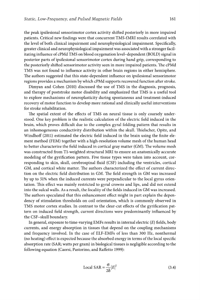
following equation (Caorsi, Pastorino, and Raffetto 1999):
Local SAR =
σ E
2
(3.4)
2δ
Static, Low-Frequency, and Pulsed Magnetic Fields
the peak ipsilesional sensorimotor cortex activity shifted posteriorly in more impaired
patients. Critical new findings were that concurrent TMS-fMRI results correlated with
the level of both clinical impairment and neurophysiological impairment. Specifically,
greater clinical and neurophysiological impairment was associated with a stronger facilitating influence of cPMd TMS on blood oxygenation level–dependent (BOLD) signal in
posterior parts of ipsilesional sensorimotor cortex during hand grip, corresponding to
the posteriorly shifted sensorimotor activity seen in more impaired patients. The cPMd
TMS was not found to influence activity in other brain regions in either hemisphere.
The authors suggested that this state-dependent influence on ipsilesional sensorimotor
regions provides a mechanism by which cPMd supports recovered function after stroke.
Dimyan and Cohen (2010) discussed the use of TMS in the diagnosis, prognosis,
and therapy of poststroke motor disability and emphasized that TMS is a useful tool
to explore mechanisms of neuroplasticity during spontaneous and treatment-induced
recovery of motor function to develop more rational and clinically useful interventions
for stroke rehabilitation.
The spatial extent of the effects of TMS on neural tissue is only coarsely understood. One key problem is the realistic calculation of the electric field induced in the
brain, which proves difficult due to the complex gyral folding pattern that results in
an inhomogeneous conductivity distribution within the skull. Thielscher, Opitz, and
Windhoff (2011) estimated the electric field induced in the brain using the finite element method (FEM) together with a high-resolution volume mesh of the human head
to better characterize the field induced in cortical gray matter (GM). The volume mesh
was constructed from T1-weighted structural MRI to ensure an anatomically accurate
modeling of the gyrification pattern. Five tissue types were taken into account, corresponding to skin, skull, cerebrospinal fluid (CSF) including the ventricles, cortical
GM, and cortical white matter. The authors characterized the effect of current direction on the electric field distribution in GM. The field strength in GM was increased
by up to 51% when the induced currents were perpendicular to the local gyrus orientation. This effect was mainly restricted to gyral crowns and lips, and did not extend
into the sulcal walls. As a result, the focality of the fields induced in GM was increased.
The authors speculated that this enhancement effect might in part explain the dependency of stimulation thresholds on coil orientation, which is commonly observed in
TMS motor cortex studies. In contrast to the clear-cut effects of the gyrification pattern on induced field strength, current directions were predominantly influenced by
the CSF–skull boundary.
In general, exposure to time-varying EMFs results in internal electric (E) fields, body
currents, and energy absorption in tissues that depend on the coupling mechanisms
and frequency involved. In the case of ELF-EMFs of less than 300 Hz, nonthermal
(no heating) effect is expected because the absorbed energy in terms of the local specific
absorption rate (SAR; watts per gram) in biological tissues is negligible according to the
following equation (Caorsi, Pastorino, and Raffetto 1999):
Local SAR =
σ E
2
(3.4)
2δ