

147
Static, Low-Frequency, and Pulsed Magnetic Fields
conditions (3–10 T, 0–41.7 T/m) for 0.5 and 1 hours. Exposure to an SMF with a high
magnetic gradient for 1 hour significantly increased insulin secretion and insulin-1
mRNA expression. Exposure to an SMF with a high magnetic flux density for 0.5 hour
significantly enhanced responsiveness to glucose stimulation. The SMF did not affect
insulin content, cell proliferation, or cell number. These results suggest that MRI systems with higher magnetic flux densities might not cause cell proliferative or functional
damages on insulin-secreting cells.
Regarding the effects of ELF-EMF on glucose-metabolic pathways, the same research
group investigated the effects of an ELF-EMF (60 Hz, 5 mT) on β-cell survival and function in HIT-T15 cells (Sakurai et al. 2008). The authors found that exposure to ELFEMF for 5 days without glucose increased cell number, exposure for 2 days without
glucose and for 5 days with 100-mg/dL glucose increased insulin secretion to the culture medium, and exposure for 2 and 5 days with 40- and 100-mg/dL glucose increased
intracellular insulin concentration. The authors speculated that the increase in cell
number under apoptotic culture conditions by exposure to ELF-EMF could lead to new
therapeutic concepts in the treatment of diabetes.
Gaetani et al. (2009) assessed the hypothesis that exposure of human cardiospheres
(CSs) and cardiosphere-derived cells (CDCs) to an ELF-EMF, tuned at Ca 2+ - ICR, may
drive their differentiation toward a cardiac-specific phenotype.
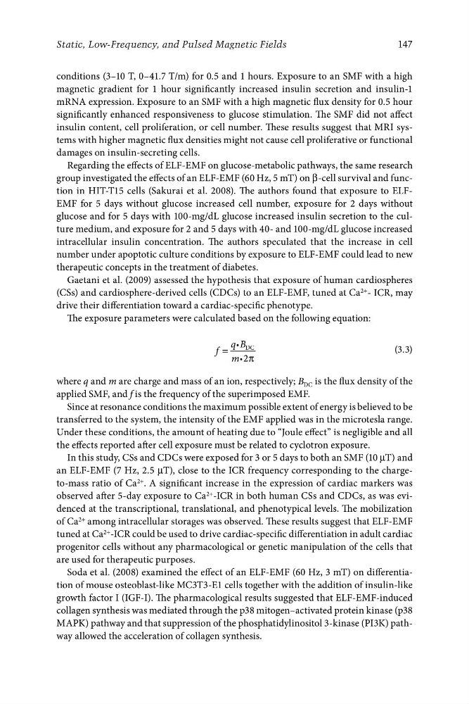
The exposure parameters were calculated based on the following equation:
q B DC
f =
i
(3.3)
mi2π
where q and m are charge and mass of an ion, respectively; B DC is the flux density of the
applied SMF, and f is the frequency of the superimposed EMF.
Since at resonance conditions the maximum possible extent of energy is believed to be
transferred to the system, the intensity of the EMF applied was in the microtesla range.
Under these conditions, the amount of heating due to “Joule effect” is negligible and all
the effects reported after cell exposure must be related to cyclotron exposure.
In this study, CSs and CDCs were exposed for 3 or 5 days to both an SMF (10 μT) and
an ELF-EMF (7 Hz, 2.5 μT), close to the ICR frequency corresponding to the chargeto-mass ratio of Ca 2+ . A significant increase in the expression of cardiac markers was
observed after 5-day exposure to Ca 2+ -ICR in both human CSs and CDCs, as was evidenced at the transcriptional, translational, and phenotypical levels. The mobilization
of Ca 2+ among intracellular storages was observed. These results suggest that ELF-EMF
tuned at Ca 2+ -ICR could be used to drive cardiac-specific differentiation in adult cardiac
progenitor cells without any pharmacological or genetic manipulation of the cells that
are used for therapeutic purposes.
Soda et al. (2008) examined the effect of an ELF-EMF (60 Hz, 3 mT) on differentiation of mouse osteoblast-like MC3T3-E1 cells together with the addition of insulin-like
growth factor I (IGF-I). The pharmacological results suggested that ELF-EMF-induced
collagen synthesis was mediated through the p38 mitogen–activated protein kinase (p38
MAPK) pathway and that suppression of the phosphatidylinositol 3-kinase (PI3K) pathway allowed the acceleration of collagen synthesis.
Static, Low-Frequency, and Pulsed Magnetic Fields
conditions (3–10 T, 0–41.7 T/m) for 0.5 and 1 hours. Exposure to an SMF with a high
magnetic gradient for 1 hour significantly increased insulin secretion and insulin-1
mRNA expression. Exposure to an SMF with a high magnetic flux density for 0.5 hour
significantly enhanced responsiveness to glucose stimulation. The SMF did not affect
insulin content, cell proliferation, or cell number. These results suggest that MRI systems with higher magnetic flux densities might not cause cell proliferative or functional
damages on insulin-secreting cells.
Regarding the effects of ELF-EMF on glucose-metabolic pathways, the same research
group investigated the effects of an ELF-EMF (60 Hz, 5 mT) on β-cell survival and function in HIT-T15 cells (Sakurai et al. 2008). The authors found that exposure to ELFEMF for 5 days without glucose increased cell number, exposure for 2 days without
glucose and for 5 days with 100-mg/dL glucose increased insulin secretion to the culture medium, and exposure for 2 and 5 days with 40- and 100-mg/dL glucose increased
intracellular insulin concentration. The authors speculated that the increase in cell
number under apoptotic culture conditions by exposure to ELF-EMF could lead to new
therapeutic concepts in the treatment of diabetes.
Gaetani et al. (2009) assessed the hypothesis that exposure of human cardiospheres
(CSs) and cardiosphere-derived cells (CDCs) to an ELF-EMF, tuned at Ca 2+ - ICR, may
drive their differentiation toward a cardiac-specific phenotype.
The exposure parameters were calculated based on the following equation:
q B DC
f =
i
(3.3)
mi2π
where q and m are charge and mass of an ion, respectively; B DC is the flux density of the
applied SMF, and f is the frequency of the superimposed EMF.
Since at resonance conditions the maximum possible extent of energy is believed to be
transferred to the system, the intensity of the EMF applied was in the microtesla range.
Under these conditions, the amount of heating due to “Joule effect” is negligible and all
the effects reported after cell exposure must be related to cyclotron exposure.
In this study, CSs and CDCs were exposed for 3 or 5 days to both an SMF (10 μT) and
an ELF-EMF (7 Hz, 2.5 μT), close to the ICR frequency corresponding to the chargeto-mass ratio of Ca 2+ . A significant increase in the expression of cardiac markers was
observed after 5-day exposure to Ca 2+ -ICR in both human CSs and CDCs, as was evidenced at the transcriptional, translational, and phenotypical levels. The mobilization
of Ca 2+ among intracellular storages was observed. These results suggest that ELF-EMF
tuned at Ca 2+ -ICR could be used to drive cardiac-specific differentiation in adult cardiac
progenitor cells without any pharmacological or genetic manipulation of the cells that
are used for therapeutic purposes.
Soda et al. (2008) examined the effect of an ELF-EMF (60 Hz, 3 mT) on differentiation of mouse osteoblast-like MC3T3-E1 cells together with the addition of insulin-like
growth factor I (IGF-I). The pharmacological results suggested that ELF-EMF-induced
collagen synthesis was mediated through the p38 mitogen–activated protein kinase (p38
MAPK) pathway and that suppression of the phosphatidylinositol 3-kinase (PI3K) pathway allowed the acceleration of collagen synthesis.