

3.1 Image Formation by Transmission Electron Microscopy (TEM)
51
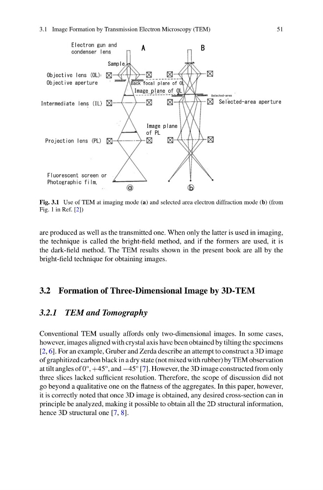
Fig. 3.1 Use of TEM at imaging mode (a) and selected area electron diffraction mode (b) (from
Fig. 1 in Ref. [2])
are produced as well as the transmitted one. When only the latter is used in imaging,
the technique is called the bright-field method, and if the formers are used, it is
the dark-field method. The TEM results shown in the present book are all by the
bright-field technique for obtaining images.
3.2 Formation of Three-Dimensional Image by 3D-TEM
3.2.1 TEM and Tomography
Conventional TEM usually affords only two-dimensional images. In some cases,
however, images aligned with crystal axis have been obtained by tilting the specimens
[2, 6]. For an example, Gruber and Zerda describe an attempt to construct a 3D image
of graphitized carbon black in a dry state (not mixed with rubber) by TEM observation
at tilt angles of 0°, +45°, and −45° [7]. However, the 3D image constructed from only
three slices lacked sufficient resolution. Therefore, the scope of discussion did not
go beyond a qualitative one on the flatness of the aggregates. In this paper, however,
it is correctly noted that once 3D image is obtained, any desired cross-section can in
principle be analyzed, making it possible to obtain all the 2D structural information,
hence 3D structural one [7, 8].
51
Fig. 3.1 Use of TEM at imaging mode (a) and selected area electron diffraction mode (b) (from
Fig. 1 in Ref. [2])
are produced as well as the transmitted one. When only the latter is used in imaging,
the technique is called the bright-field method, and if the formers are used, it is
the dark-field method. The TEM results shown in the present book are all by the
bright-field technique for obtaining images.
3.2 Formation of Three-Dimensional Image by 3D-TEM
3.2.1 TEM and Tomography
Conventional TEM usually affords only two-dimensional images. In some cases,
however, images aligned with crystal axis have been obtained by tilting the specimens
[2, 6]. For an example, Gruber and Zerda describe an attempt to construct a 3D image
of graphitized carbon black in a dry state (not mixed with rubber) by TEM observation
at tilt angles of 0°, +45°, and −45° [7]. However, the 3D image constructed from only
three slices lacked sufficient resolution. Therefore, the scope of discussion did not
go beyond a qualitative one on the flatness of the aggregates. In this paper, however,
it is correctly noted that once 3D image is obtained, any desired cross-section can in
principle be analyzed, making it possible to obtain all the 2D structural information,
hence 3D structural one [7, 8].