

1 3
Topics in Current Chemistry (2019) 377:4
3 Characterization of Carbon‑Based Single‑Atom Metal Catalysts
Characterization techniques are of great significance for studying carbon-based
single-atom metal catalysts. Two main types of technique have been widely
used to characterize these materials. One is electron microscopy and the other is
spectroscopy.
3.1 Electron Microscopy Techniques
Scanning tunneling microscopy (STM) is used for imaging of surfaces at the atomic
level. When a conducting tip approaches the surface of a sample and a bias voltage
is applied between the two, a tunneling current is generated. By monitoring the current as the tip scans across the surface, we can acquire the surface information of
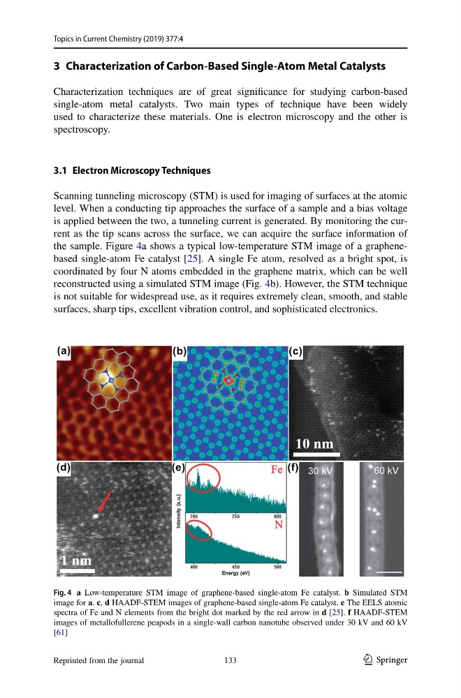
the sample. Figure 4a shows a typical low-temperature STM image of a graphenebased single-atom Fe catalyst [25]. A single Fe atom, resolved as a bright spot, is
coordinated by four N atoms embedded in the graphene matrix, which can be well
reconstructed using a simulated STM image (Fig. 4b). However, the STM technique
is not suitable for widespread use, as it requires extremely clean, smooth, and stable
surfaces, sharp tips, excellent vibration control, and sophisticated electronics.
Fig. 4 a Low-temperature STM image of graphene-based single-atom Fe catalyst. b Simulated STM
image for a. c, d HAADF-STEM images of graphene-based single-atom Fe catalyst. e The EELS atomic
spectra of Fe and N elements from the bright dot marked by the red arrow in d [25]. f HAADF-STEM
images of metallofullerene peapods in a single-wall carbon nanotube observed under 30 kV and 60 kV
[61]
Reprinted from the journal
133
Topics in Current Chemistry (2019) 377:4
3 Characterization of Carbon‑Based Single‑Atom Metal Catalysts
Characterization techniques are of great significance for studying carbon-based
single-atom metal catalysts. Two main types of technique have been widely
used to characterize these materials. One is electron microscopy and the other is
spectroscopy.
3.1 Electron Microscopy Techniques
Scanning tunneling microscopy (STM) is used for imaging of surfaces at the atomic
level. When a conducting tip approaches the surface of a sample and a bias voltage
is applied between the two, a tunneling current is generated. By monitoring the current as the tip scans across the surface, we can acquire the surface information of
the sample. Figure 4a shows a typical low-temperature STM image of a graphenebased single-atom Fe catalyst [25]. A single Fe atom, resolved as a bright spot, is
coordinated by four N atoms embedded in the graphene matrix, which can be well
reconstructed using a simulated STM image (Fig. 4b). However, the STM technique
is not suitable for widespread use, as it requires extremely clean, smooth, and stable
surfaces, sharp tips, excellent vibration control, and sophisticated electronics.
Fig. 4 a Low-temperature STM image of graphene-based single-atom Fe catalyst. b Simulated STM
image for a. c, d HAADF-STEM images of graphene-based single-atom Fe catalyst. e The EELS atomic
spectra of Fe and N elements from the bright dot marked by the red arrow in d [25]. f HAADF-STEM
images of metallofullerene peapods in a single-wall carbon nanotube observed under 30 kV and 60 kV
[61]
Reprinted from the journal
133