

430
8 Morphogenesis
of the beam to bend concave upward, but the curvature near the left end reverses
to meet the zero-slope condition at the fixed end. As the growth region shrinks
and becomes more localized near the left end, the plate near that end begins to
bend upward like a cantilever plate with a growing lower layer (see Fig. 8.12a). The
constraint at the right end forces the beam outside this region to bend in the opposite
direction, reversing the overall curvature of the plate.
Now, we turn to some problems involving morphogenesis in real embryos.
8.4 Gastrulation
As discussed in Sect. 1.4.2, the fertilized egg divides into cells that form a hollow,
fluid-filled epithelial shell called the blastula. Then, during gastrulation, the cells
in the blastula become organized into three germ layers, which later create specific
tissues and organs. The mechanisms driving this process are species dependent, but
all involve cells moving from the outer shell into the fluid-filled cavity, or blastocoel.
The cells remaining in the outer layer form ectoderm, while those moving inside
form endoderm (inner layer) and mesoderm (middle layer) (see Fig. 1.2).
Cells enter the blastocoel in two main ways. Either the outer layer invaginates
(folds inward), or epithelial cells break free of their attachments and crawl inside as
mesenchymal cells. Here, we consider the fundamental mechanics of invagination,
followed by examples from a few well-studied species.
8.4.1 Invagination Mechanics
In most cases, invagination is driven by active bending. (Evagination, i.e., outward
bending, also occurs but is far less common.) As discussed earlier in this chapter,
bending can be caused by differential growth or contraction across the epithelial
thickness. The shape and relative size of the invagination depend on the spatial
extent of the driving forces, the geometry of the epithelium, and any external
constraints.
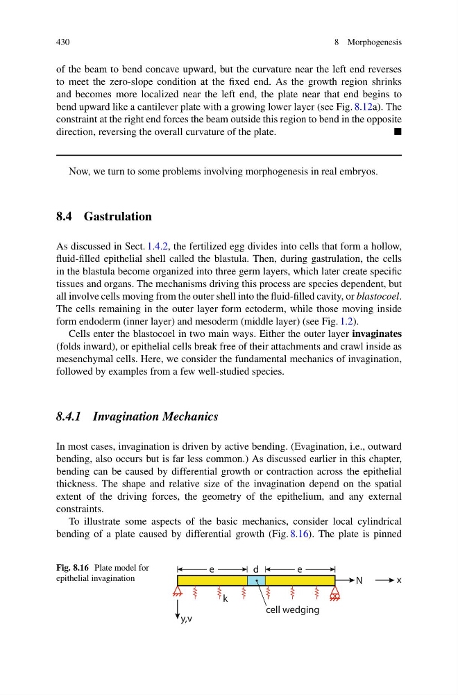
To illustrate some aspects of the basic mechanics, consider local cylindrical
bending of a plate caused by differential growth (Fig. 8.16). The plate is pinned
Fig. 8.16 Plate model for
epithelial invagination
x
y,v
cell wedging
d
N
k
e
e
8 Morphogenesis
of the beam to bend concave upward, but the curvature near the left end reverses
to meet the zero-slope condition at the fixed end. As the growth region shrinks
and becomes more localized near the left end, the plate near that end begins to
bend upward like a cantilever plate with a growing lower layer (see Fig. 8.12a). The
constraint at the right end forces the beam outside this region to bend in the opposite
direction, reversing the overall curvature of the plate.
Now, we turn to some problems involving morphogenesis in real embryos.
8.4 Gastrulation
As discussed in Sect. 1.4.2, the fertilized egg divides into cells that form a hollow,
fluid-filled epithelial shell called the blastula. Then, during gastrulation, the cells
in the blastula become organized into three germ layers, which later create specific
tissues and organs. The mechanisms driving this process are species dependent, but
all involve cells moving from the outer shell into the fluid-filled cavity, or blastocoel.
The cells remaining in the outer layer form ectoderm, while those moving inside
form endoderm (inner layer) and mesoderm (middle layer) (see Fig. 1.2).
Cells enter the blastocoel in two main ways. Either the outer layer invaginates
(folds inward), or epithelial cells break free of their attachments and crawl inside as
mesenchymal cells. Here, we consider the fundamental mechanics of invagination,
followed by examples from a few well-studied species.
8.4.1 Invagination Mechanics
In most cases, invagination is driven by active bending. (Evagination, i.e., outward
bending, also occurs but is far less common.) As discussed earlier in this chapter,
bending can be caused by differential growth or contraction across the epithelial
thickness. The shape and relative size of the invagination depend on the spatial
extent of the driving forces, the geometry of the epithelium, and any external
constraints.
To illustrate some aspects of the basic mechanics, consider local cylindrical
bending of a plate caused by differential growth (Fig. 8.16). The plate is pinned
Fig. 8.16 Plate model for
epithelial invagination
x
y,v
cell wedging
d
N
k
e
e