

10
1 Introduction
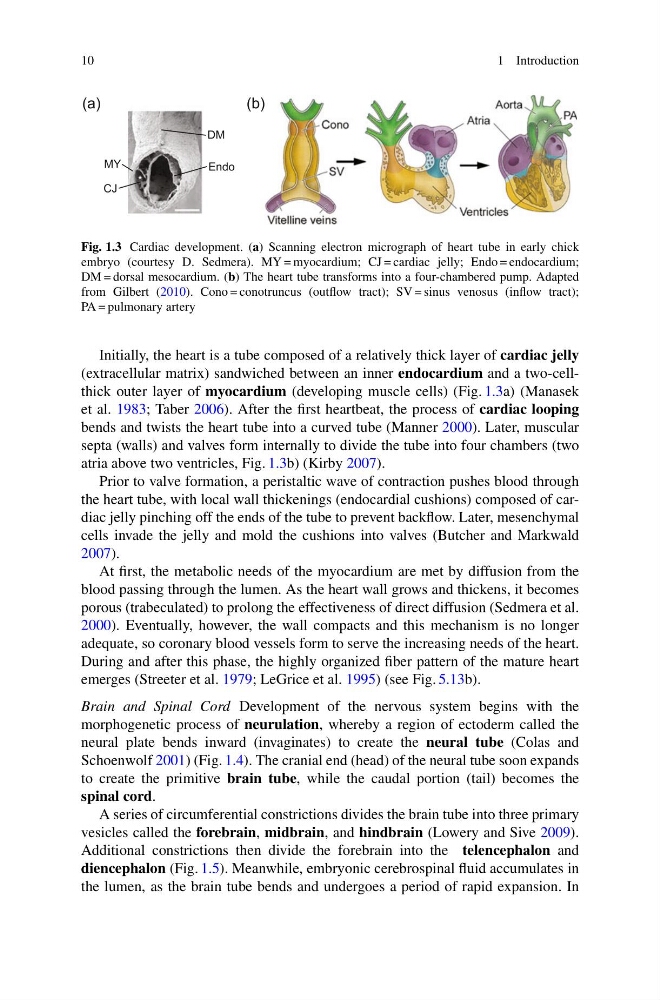
Fig. 1.3 Cardiac development. (a) Scanning electron micrograph of heart tube in early chick
embryo (courtesy D. Sedmera). MY = myocardium; CJ = cardiac jelly; Endo = endocardium;
DM = dorsal mesocardium. (b) The heart tube transforms into a four-chambered pump. Adapted
from Gilbert (2010). Cono = conotruncus (outflow tract); SV = sinus venosus (inflow tract);
PA = pulmonary artery
Initially, the heart is a tube composed of a relatively thick layer of cardiac jelly
(extracellular matrix) sandwiched between an inner endocardium and a two-cellthick outer layer of myocardium (developing muscle cells) (Fig. 1.3a) (Manasek
et al. 1983; Taber 2006). After the first heartbeat, the process of cardiac looping
bends and twists the heart tube into a curved tube (Manner 2000). Later, muscular
septa (walls) and valves form internally to divide the tube into four chambers (two
atria above two ventricles, Fig. 1.3b) (Kirby 2007).
Prior to valve formation, a peristaltic wave of contraction pushes blood through
the heart tube, with local wall thickenings (endocardial cushions) composed of cardiac jelly pinching off the ends of the tube to prevent backflow. Later, mesenchymal
cells invade the jelly and mold the cushions into valves (Butcher and Markwald
2007).
At first, the metabolic needs of the myocardium are met by diffusion from the
blood passing through the lumen. As the heart wall grows and thickens, it becomes
porous (trabeculated) to prolong the effectiveness of direct diffusion (Sedmera et al.
2000). Eventually, however, the wall compacts and this mechanism is no longer
adequate, so coronary blood vessels form to serve the increasing needs of the heart.
During and after this phase, the highly organized fiber pattern of the mature heart
emerges (Streeter et al. 1979; LeGrice et al. 1995) (see Fig. 5.13b).
Brain and Spinal Cord Development of the nervous system begins with the
morphogenetic process of neurulation, whereby a region of ectoderm called the
neural plate bends inward (invaginates) to create the neural tube (Colas and
Schoenwolf 2001) (Fig. 1.4). The cranial end (head) of the neural tube soon expands
to create the primitive brain tube, while the caudal portion (tail) becomes the
spinal cord.
A series of circumferential constrictions divides the brain tube into three primary
vesicles called the forebrain, midbrain, and hindbrain (Lowery and Sive 2009).
Additional constrictions then divide the forebrain into the telencephalon and
diencephalon (Fig. 1.5). Meanwhile, embryonic cerebrospinal fluid accumulates in
the lumen, as the brain tube bends and undergoes a period of rapid expansion. In
1 Introduction
Fig. 1.3 Cardiac development. (a) Scanning electron micrograph of heart tube in early chick
embryo (courtesy D. Sedmera). MY = myocardium; CJ = cardiac jelly; Endo = endocardium;
DM = dorsal mesocardium. (b) The heart tube transforms into a four-chambered pump. Adapted
from Gilbert (2010). Cono = conotruncus (outflow tract); SV = sinus venosus (inflow tract);
PA = pulmonary artery
Initially, the heart is a tube composed of a relatively thick layer of cardiac jelly
(extracellular matrix) sandwiched between an inner endocardium and a two-cellthick outer layer of myocardium (developing muscle cells) (Fig. 1.3a) (Manasek
et al. 1983; Taber 2006). After the first heartbeat, the process of cardiac looping
bends and twists the heart tube into a curved tube (Manner 2000). Later, muscular
septa (walls) and valves form internally to divide the tube into four chambers (two
atria above two ventricles, Fig. 1.3b) (Kirby 2007).
Prior to valve formation, a peristaltic wave of contraction pushes blood through
the heart tube, with local wall thickenings (endocardial cushions) composed of cardiac jelly pinching off the ends of the tube to prevent backflow. Later, mesenchymal
cells invade the jelly and mold the cushions into valves (Butcher and Markwald
2007).
At first, the metabolic needs of the myocardium are met by diffusion from the
blood passing through the lumen. As the heart wall grows and thickens, it becomes
porous (trabeculated) to prolong the effectiveness of direct diffusion (Sedmera et al.
2000). Eventually, however, the wall compacts and this mechanism is no longer
adequate, so coronary blood vessels form to serve the increasing needs of the heart.
During and after this phase, the highly organized fiber pattern of the mature heart
emerges (Streeter et al. 1979; LeGrice et al. 1995) (see Fig. 5.13b).
Brain and Spinal Cord Development of the nervous system begins with the
morphogenetic process of neurulation, whereby a region of ectoderm called the
neural plate bends inward (invaginates) to create the neural tube (Colas and
Schoenwolf 2001) (Fig. 1.4). The cranial end (head) of the neural tube soon expands
to create the primitive brain tube, while the caudal portion (tail) becomes the
spinal cord.
A series of circumferential constrictions divides the brain tube into three primary
vesicles called the forebrain, midbrain, and hindbrain (Lowery and Sive 2009).
Additional constrictions then divide the forebrain into the telencephalon and
diencephalon (Fig. 1.5). Meanwhile, embryonic cerebrospinal fluid accumulates in
the lumen, as the brain tube bends and undergoes a period of rapid expansion. In