

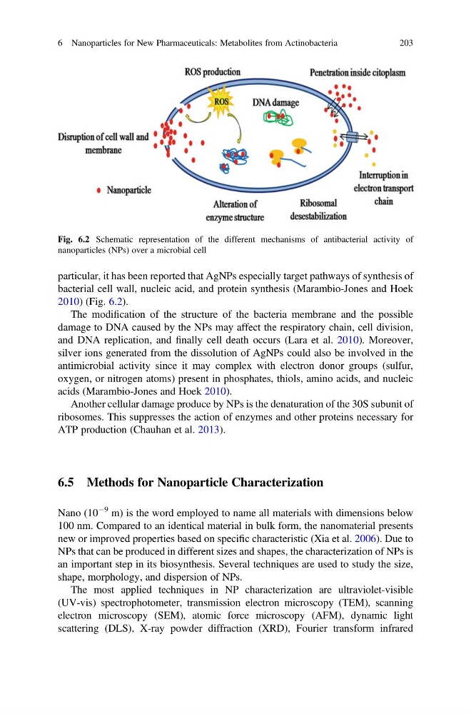
particular, it has been reported that AgNPs especially target pathways of synthesis of
bacterial cell wall, nucleic acid, and protein synthesis (Marambio-Jones and Hoek
2010) (Fig. 6.2).
The modification of the structure of the bacteria membrane and the possible
damage to DNA caused by the NPs may affect the respiratory chain, cell division,
and DNA replication, and finally cell death occurs (Lara et al. 2010). Moreover,
silver ions generated from the dissolution of AgNPs could also be involved in the
antimicrobial activity since it may complex with electron donor groups (sulfur,
oxygen, or nitrogen atoms) present in phosphates, thiols, amino acids, and nucleic
acids (Marambio-Jones and Hoek 2010).
Another cellular damage produce by NPs is the denaturation of the 30S subunit of
ribosomes. This suppresses the action of enzymes and other proteins necessary for
ATP production (Chauhan et al. 2013).
6.5 Methods for Nanoparticle Characterization
Nano (10
À9 m) is the word employed to name all materials with dimensions below
100 nm. Compared to an identical material in bulk form, the nanomaterial presents
new or improved properties based on specific characteristic (Xia et al. 2006). Due to
NPs that can be produced in different sizes and shapes, the characterization of NPs is
an important step in its biosynthesis. Several techniques are used to study the size,
shape, morphology, and dispersion of NPs.
The most applied techniques in NP characterization are ultraviolet-visible
(UV-vis) spectrophotometer, transmission electron microscopy (TEM), scanning
electron microscopy (SEM), atomic force microscopy (AFM), dynamic light
scattering (DLS), X-ray powder diffraction (XRD), Fourier transform infrared
Fig. 6.2 Schematic representation of the different mechanisms of antibacterial activity of
nanoparticles (NPs) over a microbial cell
6 Nanoparticles for New Pharmaceuticals: Metabolites from Actinobacteria
203
bacterial cell wall, nucleic acid, and protein synthesis (Marambio-Jones and Hoek
2010) (Fig. 6.2).
The modification of the structure of the bacteria membrane and the possible
damage to DNA caused by the NPs may affect the respiratory chain, cell division,
and DNA replication, and finally cell death occurs (Lara et al. 2010). Moreover,
silver ions generated from the dissolution of AgNPs could also be involved in the
antimicrobial activity since it may complex with electron donor groups (sulfur,
oxygen, or nitrogen atoms) present in phosphates, thiols, amino acids, and nucleic
acids (Marambio-Jones and Hoek 2010).
Another cellular damage produce by NPs is the denaturation of the 30S subunit of
ribosomes. This suppresses the action of enzymes and other proteins necessary for
ATP production (Chauhan et al. 2013).
6.5 Methods for Nanoparticle Characterization
Nano (10
À9 m) is the word employed to name all materials with dimensions below
100 nm. Compared to an identical material in bulk form, the nanomaterial presents
new or improved properties based on specific characteristic (Xia et al. 2006). Due to
NPs that can be produced in different sizes and shapes, the characterization of NPs is
an important step in its biosynthesis. Several techniques are used to study the size,
shape, morphology, and dispersion of NPs.
The most applied techniques in NP characterization are ultraviolet-visible
(UV-vis) spectrophotometer, transmission electron microscopy (TEM), scanning
electron microscopy (SEM), atomic force microscopy (AFM), dynamic light
scattering (DLS), X-ray powder diffraction (XRD), Fourier transform infrared
Fig. 6.2 Schematic representation of the different mechanisms of antibacterial activity of
nanoparticles (NPs) over a microbial cell
6 Nanoparticles for New Pharmaceuticals: Metabolites from Actinobacteria
203