

drugs or fluorescence dyes were often integrated into the assemblies, generating
multifunctional and multi-stimuli-responsive systems for drug delivery, magnetic
resonance imaging (MRI), and magnetofection.
20.3.2 One-Dimensional Magnetic Supramolecular Nanofibers
In the nature, one-dimensional magnetic supramolecular assembly is a well-known
phenomenon; in the magnetotactic bacteria, the magnetosomes could self-assemble to
form one-dimensional nanofibers [24, 25]. In the biomimetic area, it is very interesting
to mimic the formation of magnetosome-composed nanofibers by controlling the
supramolecular interaction between the magnetic nanoparticles. Recently, we
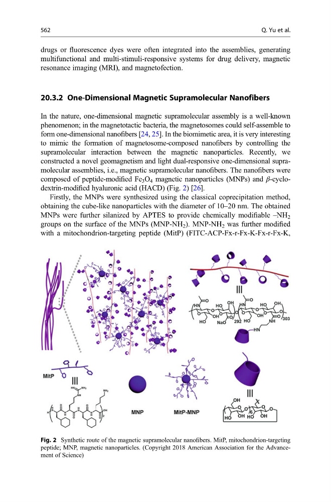
constructed a novel geomagnetism and light dual-responsive one-dimensional supramolecular assemblies, i.e., magnetic supramolecular nanofibers. The nanofibers were
composed of peptide-modified Fe 3 O 4 magnetic nanoparticles (MNPs) and β-cyclodextrin-modified hyaluronic acid (HACD) (Fig. 2) [26].
Firstly, the MNPs were synthesized using the classical coprecipitation method,
obtaining the cube-like nanoparticles with the diameter of 10–20 nm. The obtained
MNPs were further silanized by APTES to provide chemically modifiable –NH 2
groups on the surface of the MNPs (MNP-NH 2 ). MNP-NH 2 was further modified
with a mitochondrion-targeting peptide (MitP) (FITC-ACP-Fx-r-Fx-K-Fx-r-Fx-K,
Fig. 2 Synthetic route of the magnetic supramolecular nanofibers. MitP, mitochondrion-targeting
peptide; MNP, magnetic nanoparticles. (Copyright 2018 American Association for the Advancement of Science)
562
Q. Yu et al.