

prominent advantage of stimuli-responsive delivery systems is precise controlled
release as a result of specific stimuli.
Jia and coworkers [74] developed cyclodextrin vesicles (CDVs) for capture and
release of myoglobin (Myo), which is a very useful cardiac biomarker for early
detection of acute myocardial infarction [72, 73]. These authors prepared a captureand-release system, including so-called SH-CDVs, which were grafted onto a
mesoporous poly(glycidyl methacrylate–pentaerythritol triacrylate) (poly
(GMA-PETA)) monolith, and SH-CDVs was the key factor in this pH-reversible
system. The SH-CDVs (diameter ~90 nm) presents as vesicle shapes when the
environmental pH is 7.4, but when the pH is decreased to 5.0, the vesicle shapes
transform into fiber tubes (diameter ~8 nm) (Fig. 16a). On the basis of this fact, the
authors loaded the myoglobin into the sample when the SH-CD was in the form of
tubes, and then they increased the buffer pH to 7.4, causing the SH-CD to transform
into vesicle shapes and uptake the myoglobin at the same time. The mixture was then
washed to remove other molecules from the vesicles. Finally, the buffer was acidified
to 5.0 in order to release the myoglobin, which could be detected by mass spectrometry and other forms of detection [74] (Fig. 16b).
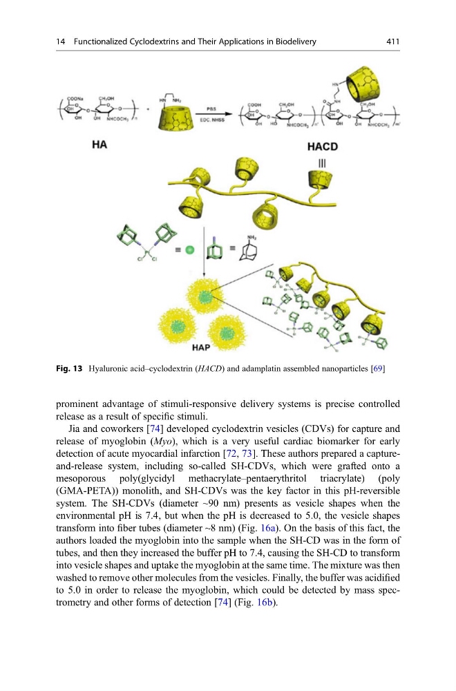
Fig. 13 Hyaluronic acid–cyclodextrin (HACD) and adamplatin assembled nanoparticles [69]
14 Functionalized Cyclodextrins and Their Applications in Biodelivery
411
release as a result of specific stimuli.
Jia and coworkers [74] developed cyclodextrin vesicles (CDVs) for capture and
release of myoglobin (Myo), which is a very useful cardiac biomarker for early
detection of acute myocardial infarction [72, 73]. These authors prepared a captureand-release system, including so-called SH-CDVs, which were grafted onto a
mesoporous poly(glycidyl methacrylate–pentaerythritol triacrylate) (poly
(GMA-PETA)) monolith, and SH-CDVs was the key factor in this pH-reversible
system. The SH-CDVs (diameter ~90 nm) presents as vesicle shapes when the
environmental pH is 7.4, but when the pH is decreased to 5.0, the vesicle shapes
transform into fiber tubes (diameter ~8 nm) (Fig. 16a). On the basis of this fact, the
authors loaded the myoglobin into the sample when the SH-CD was in the form of
tubes, and then they increased the buffer pH to 7.4, causing the SH-CD to transform
into vesicle shapes and uptake the myoglobin at the same time. The mixture was then
washed to remove other molecules from the vesicles. Finally, the buffer was acidified
to 5.0 in order to release the myoglobin, which could be detected by mass spectrometry and other forms of detection [74] (Fig. 16b).
Fig. 13 Hyaluronic acid–cyclodextrin (HACD) and adamplatin assembled nanoparticles [69]
14 Functionalized Cyclodextrins and Their Applications in Biodelivery
411