

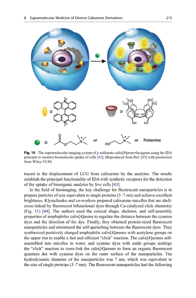
traced to the displacement of LCG from calixarene by the analytes. The results
establish the principal functionality of IDA with synthetic receptors for the detection
of the uptake of bioorganic analytes by live cells [43].
In the field of bioimaging, the key challenge for fluorescent nanoparticles is to
prepare particles of size equivalent to single proteins (3–7 nm) and achieve excellent
brightness. Klymchenko and co-workers prepared calixarene micelles that are shellcross-linked by fluorescent bifunctional dyes through Cu-catalyzed click chemistry
(Fig. 11) [44]. The authors used the conical shape, skeleton, and self-assembly
properties of amphiphiles calix[4]arene to regulate the distance between the cyanine
dyes and the direction of the dye. Finally, they obtained protein-sized fluorescent
nanoparticles and minimized the self-quenching between the fluorescent dyes. They
synthesized positively charged amphiphilic calix[4]arenes with acetylene groups on
the upper rim to enable a fast and efficient “click” reaction. The calix[4]arenes selfassembled into micelles in water, and cyanine dyes with azide groups undergo
the “click” reaction to cross-link the calix[4]arenes to form an organic fluorescent
quantum dot with cyanine dyes on the outer surface of the nanoparticles. The
hydrodynamic diameter of the nanoparticles was 7 nm, which was equivalent to
the size of single proteins (3–7 nm). The fluorescent nanoparticles had the following
Fig. 10 The supramolecular imaging system of p-sulfonato-calix[4]arene•lucigenin using the IDA
principle to monitor biomolecule uptake of cells [43]. (Reproduced from Ref. [43] with permission
from Wiley-VCH)
8 Supramolecular Medicine of Diverse Calixarene Derivatives
215
establish the principal functionality of IDA with synthetic receptors for the detection
of the uptake of bioorganic analytes by live cells [43].
In the field of bioimaging, the key challenge for fluorescent nanoparticles is to
prepare particles of size equivalent to single proteins (3–7 nm) and achieve excellent
brightness. Klymchenko and co-workers prepared calixarene micelles that are shellcross-linked by fluorescent bifunctional dyes through Cu-catalyzed click chemistry
(Fig. 11) [44]. The authors used the conical shape, skeleton, and self-assembly
properties of amphiphiles calix[4]arene to regulate the distance between the cyanine
dyes and the direction of the dye. Finally, they obtained protein-sized fluorescent
nanoparticles and minimized the self-quenching between the fluorescent dyes. They
synthesized positively charged amphiphilic calix[4]arenes with acetylene groups on
the upper rim to enable a fast and efficient “click” reaction. The calix[4]arenes selfassembled into micelles in water, and cyanine dyes with azide groups undergo
the “click” reaction to cross-link the calix[4]arenes to form an organic fluorescent
quantum dot with cyanine dyes on the outer surface of the nanoparticles. The
hydrodynamic diameter of the nanoparticles was 7 nm, which was equivalent to
the size of single proteins (3–7 nm). The fluorescent nanoparticles had the following
Fig. 10 The supramolecular imaging system of p-sulfonato-calix[4]arene•lucigenin using the IDA
principle to monitor biomolecule uptake of cells [43]. (Reproduced from Ref. [43] with permission
from Wiley-VCH)
8 Supramolecular Medicine of Diverse Calixarene Derivatives
215