

control) at a concentration of 1 μM. A more detailed investigation of binding
kinetics was provided for JAK3 vs. BTK, and it was shown that the k inact /K I ratio
was at least three orders of magnitude higher for JAK3 (k inact /K I ¼ 3.0 Â 10
5 M
À1 s
À1 )
suggesting pronounced kinetic selectivity.
Nevertheless, compound 66 failed in murine PK models revealing low oral
bioavailability and high clearance. Therefore, the same development strategy was
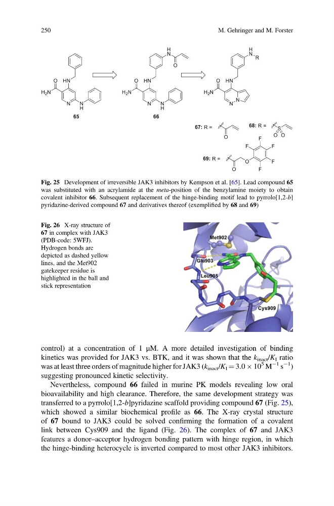
transferred to a pyrrolo[1,2-b]pyridazine scaffold providing compound 67 (Fig. 25),
which showed a similar biochemical profile as 66. The X-ray crystal structure
of 67 bound to JAK3 could be solved confirming the formation of a covalent
link between Cys909 and the ligand (Fig. 26). The complex of 67 and JAK3
features a donor–acceptor hydrogen bonding pattern with hinge region, in which
the hinge-binding heterocycle is inverted compared to most other JAK3 inhibitors.
Fig. 26 X-ray structure of
67 in complex with JAK3
(PDB-code: 5WFJ).
Hydrogen bonds are
depicted as dashed yellow
lines, and the Met902
gatekeeper residue is
highlighted in the ball and
stick representation
Fig. 25 Development of irreversible JAK3 inhibitors by Kempson et al. [65]. Lead compound 65
was substituted with an acrylamide at the meta-position of the benzylamine moiety to obtain
covalent inhibitor 66. Subsequent replacement of the hinge-binding motif lead to pyrrolo[1,2-b]
pyridazine-derived compound 67 and derivatives thereof (exemplified by 68 and 69)
250
M. Gehringer and M. Forster
kinetics was provided for JAK3 vs. BTK, and it was shown that the k inact /K I ratio
was at least three orders of magnitude higher for JAK3 (k inact /K I ¼ 3.0 Â 10
5 M
À1 s
À1 )
suggesting pronounced kinetic selectivity.
Nevertheless, compound 66 failed in murine PK models revealing low oral
bioavailability and high clearance. Therefore, the same development strategy was
transferred to a pyrrolo[1,2-b]pyridazine scaffold providing compound 67 (Fig. 25),
which showed a similar biochemical profile as 66. The X-ray crystal structure
of 67 bound to JAK3 could be solved confirming the formation of a covalent
link between Cys909 and the ligand (Fig. 26). The complex of 67 and JAK3
features a donor–acceptor hydrogen bonding pattern with hinge region, in which
the hinge-binding heterocycle is inverted compared to most other JAK3 inhibitors.
Fig. 26 X-ray structure of
67 in complex with JAK3
(PDB-code: 5WFJ).
Hydrogen bonds are
depicted as dashed yellow
lines, and the Met902
gatekeeper residue is
highlighted in the ball and
stick representation
Fig. 25 Development of irreversible JAK3 inhibitors by Kempson et al. [65]. Lead compound 65
was substituted with an acrylamide at the meta-position of the benzylamine moiety to obtain
covalent inhibitor 66. Subsequent replacement of the hinge-binding motif lead to pyrrolo[1,2-b]
pyridazine-derived compound 67 and derivatives thereof (exemplified by 68 and 69)
250
M. Gehringer and M. Forster