

Whereas JNK1 and JNK2 are present in all cells and tissues of the human
body, JNK3 is almost exclusively expressed in the brain and to a lesser extent
in the testis and heart. The deregulation of JNK3 is linked to several disorders
ranging from neurodegenerative diseases, like Parkinson’s and Alzheimer’s disease,
to inflammation, metabolic diseases, diabetes, liver diseases, and cancer [6–8].
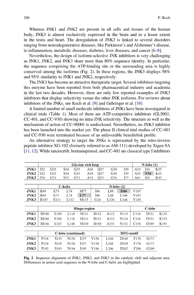
Nevertheless, the design of isoform-selective JNK inhibitors is very challenging
as JNK1, JNK2, and JNK3 share more than 80% sequence identity. In particular,
the sequence comprising the ATP-binding site or the surrounding area is highly
conserved among the isoforms (Fig. 2). In these regions, the JNK3 displays 98%
and 95% similarity to JNK1 and JNK2, respectively.
The JNK3 has become an attractive therapeutic target. Several inhibitors targeting
this enzyme have been reported from both pharmaceutical industry and academia
in the last two decades. However, there are only few reported examples of JNK3
inhibitors that display selectivity versus the other JNK isoforms. For reviews about
inhibitors of the JNKs, see Koch et al. [9] and Gehringer et al. [10]
A limited number of small molecule inhibitors of JNKs have been investigated in
clinical trials (Table 1). Most of them are ATP-competitive inhibitors (GL5001,
CC-401, and CC-930) showing no intra-JNK selectivity. The structure as well as the
mechanism of action of CC-90001 is undisclosed. Nevertheless, no JNK3 inhibitor
has been launched into the market yet. The phase II clinical trial studies of CC-401
and CC-930 were terminated because of an unfavorable benefit/risk profile.
An alternative strategy to target the JNKs is represented by the retro-inverso
peptide inhibitor XG-102 (formerly referred to as AM-111) developed by Xigen SA
[11, 12]. While tanzisertib, bentamapimod, and CC-401 are classical type I inhibitors
Glycine rich loop
N-lobe (1)
JNK1 I32 G33
S34 G35
A36 Q37
G38
I39
A53 I54
K55
JNK2 I32 G33
S34 G35
A36 Q37
G38
I39
A53 V54 K55
JNK3 I70 G71
S72 G73
A74 Q75
G76
I77
A91 I92
K93
C-helix
N-lobe (2)
JNK1 R69
E73
L74
M77
I86
L88
I106
V107
JNK2 R69
E73
L74
L77
I86
L88
L106
V107
JNK3 R107
E111
L112
M115
I124
L126
L144
V145
Hinge-region
C-lobe
JNK1 M108 E109
L110 M111 D112 A113 N114 C116
D151 K153
JNK2 M108 E109
L110 M111 D112 A113 N114 C116
D151 K153
JNK3 M146 E147
L148 M149 D150 A151 N152 C154
D189 K191
C-lobe (continued)
DFG-motif
JNK1 P154
S155
N156 I157 V158
L168
D169
F170
G171
JNK2 P154
S155
N156 I157 V158
L168
D169
F170
G171
JNK3 P192
S193
N194 I195 V196
L206
D207
F208
G209
Fig. 2 Sequence alignment of JNK1, JNK2, and JNK3 in the catalytic cleft and adjacent area.
Differences in amino acid sequence in the N-lobe and C-helix are highlighted
206
P. Koch
body, JNK3 is almost exclusively expressed in the brain and to a lesser extent
in the testis and heart. The deregulation of JNK3 is linked to several disorders
ranging from neurodegenerative diseases, like Parkinson’s and Alzheimer’s disease,
to inflammation, metabolic diseases, diabetes, liver diseases, and cancer [6–8].
Nevertheless, the design of isoform-selective JNK inhibitors is very challenging
as JNK1, JNK2, and JNK3 share more than 80% sequence identity. In particular,
the sequence comprising the ATP-binding site or the surrounding area is highly
conserved among the isoforms (Fig. 2). In these regions, the JNK3 displays 98%
and 95% similarity to JNK1 and JNK2, respectively.
The JNK3 has become an attractive therapeutic target. Several inhibitors targeting
this enzyme have been reported from both pharmaceutical industry and academia
in the last two decades. However, there are only few reported examples of JNK3
inhibitors that display selectivity versus the other JNK isoforms. For reviews about
inhibitors of the JNKs, see Koch et al. [9] and Gehringer et al. [10]
A limited number of small molecule inhibitors of JNKs have been investigated in
clinical trials (Table 1). Most of them are ATP-competitive inhibitors (GL5001,
CC-401, and CC-930) showing no intra-JNK selectivity. The structure as well as the
mechanism of action of CC-90001 is undisclosed. Nevertheless, no JNK3 inhibitor
has been launched into the market yet. The phase II clinical trial studies of CC-401
and CC-930 were terminated because of an unfavorable benefit/risk profile.
An alternative strategy to target the JNKs is represented by the retro-inverso
peptide inhibitor XG-102 (formerly referred to as AM-111) developed by Xigen SA
[11, 12]. While tanzisertib, bentamapimod, and CC-401 are classical type I inhibitors
Glycine rich loop
N-lobe (1)
JNK1 I32 G33
S34 G35
A36 Q37
G38
I39
A53 I54
K55
JNK2 I32 G33
S34 G35
A36 Q37
G38
I39
A53 V54 K55
JNK3 I70 G71
S72 G73
A74 Q75
G76
I77
A91 I92
K93
C-helix
N-lobe (2)
JNK1 R69
E73
L74
M77
I86
L88
I106
V107
JNK2 R69
E73
L74
L77
I86
L88
L106
V107
JNK3 R107
E111
L112
M115
I124
L126
L144
V145
Hinge-region
C-lobe
JNK1 M108 E109
L110 M111 D112 A113 N114 C116
D151 K153
JNK2 M108 E109
L110 M111 D112 A113 N114 C116
D151 K153
JNK3 M146 E147
L148 M149 D150 A151 N152 C154
D189 K191
C-lobe (continued)
DFG-motif
JNK1 P154
S155
N156 I157 V158
L168
D169
F170
G171
JNK2 P154
S155
N156 I157 V158
L168
D169
F170
G171
JNK3 P192
S193
N194 I195 V196
L206
D207
F208
G209
Fig. 2 Sequence alignment of JNK1, JNK2, and JNK3 in the catalytic cleft and adjacent area.
Differences in amino acid sequence in the N-lobe and C-helix are highlighted
206
P. Koch