

residues. Mutating this residue to glycine, which is not present in any human
kinases, has been exploited for the development of kinase-specific ATP analogues
for functional studies [28].
Combination of two rare sequence variations led to exclusively selective
inhibitors. For instance, the p38 inhibitor skepinone-L is a potent and selective
type-I inhibitor exploiting the presence of a small gatekeeper residue and an unusual
glycine residue located in the kinase hinge region [90–92].
Canonical type-II inhibitors are ATP competitive and target an inactive state
of the kinase catalytic domain which is created by the “outward” flip of the DFG
motif. This binding mode gained popularity in drug design after it has been
found that the first approved kinase inhibitor Gleevec induces and stabilizes this
conformation in its main targets ABL and KIT kinase [93, 94]. All type-II inhibitors
protrude into the so-called deep pocket which is made accessible in this
inactive state. Type-II inhibitors are therefore slightly elongated small molecules
when compared to type-I inhibitors. However, the type-II binding mode needs
to be confirmed experimentally as a large diversity of binding modes have
been observed for putative type-II inhibitors including canonical type-I interactions.
In the DFG-out state, the aspartate of the DFG rotates ~180
and moves ~5 Å away
from the ATP-binding site, inactivating the kinase. However, the canonical DFG-out
state represents only one of many possible conformations of the DFG motif,
and a large number of intermediate states have been described.
Initial expectations that type-II inhibitors would be more selective since
they include the dynamic properties of the DFG-out movement were however
not confirmed by more comprehensive studies, demonstrating that the DFG-out
conformation can be induced by many kinases including also CDKs that are
additionally constrained by interaction with cyclins [95–98]. A number of studies
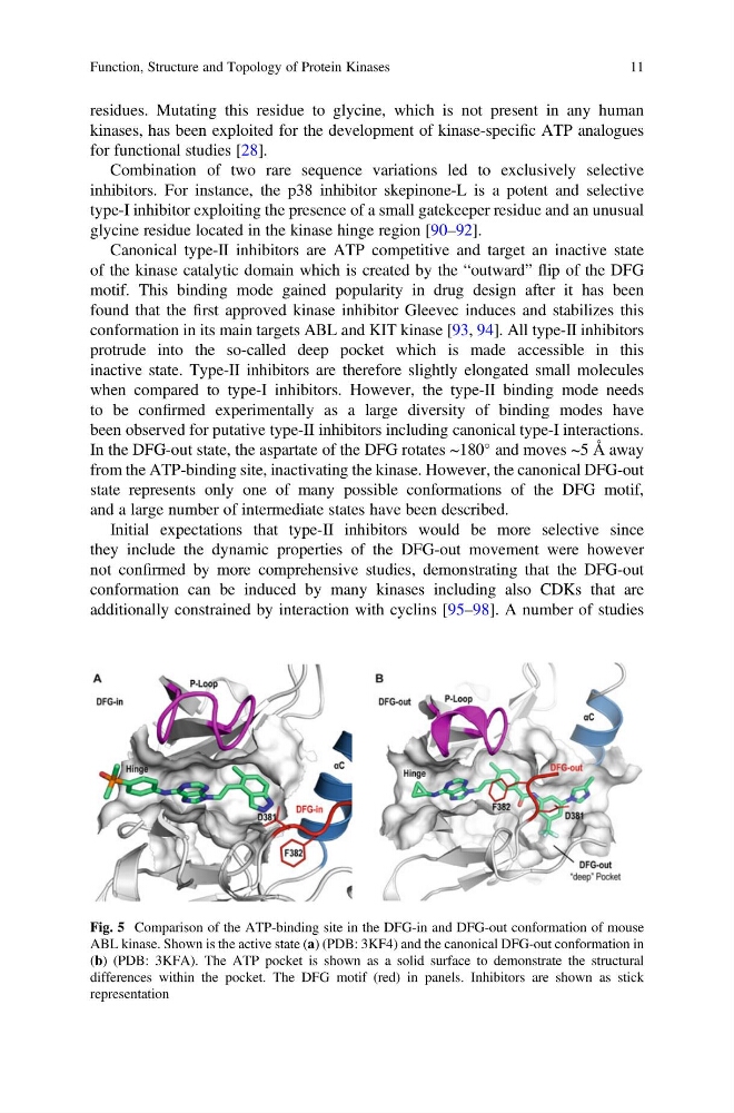
Fig. 5 Comparison of the ATP-binding site in the DFG-in and DFG-out conformation of mouse
ABL kinase. Shown is the active state (a) (PDB: 3KF4) and the canonical DFG-out conformation in
(b) (PDB: 3KFA). The ATP pocket is shown as a solid surface to demonstrate the structural
differences within the pocket. The DFG motif (red) in panels. Inhibitors are shown as stick
representation
Function, Structure and Topology of Protein Kinases
11
kinases, has been exploited for the development of kinase-specific ATP analogues
for functional studies [28].
Combination of two rare sequence variations led to exclusively selective
inhibitors. For instance, the p38 inhibitor skepinone-L is a potent and selective
type-I inhibitor exploiting the presence of a small gatekeeper residue and an unusual
glycine residue located in the kinase hinge region [90–92].
Canonical type-II inhibitors are ATP competitive and target an inactive state
of the kinase catalytic domain which is created by the “outward” flip of the DFG
motif. This binding mode gained popularity in drug design after it has been
found that the first approved kinase inhibitor Gleevec induces and stabilizes this
conformation in its main targets ABL and KIT kinase [93, 94]. All type-II inhibitors
protrude into the so-called deep pocket which is made accessible in this
inactive state. Type-II inhibitors are therefore slightly elongated small molecules
when compared to type-I inhibitors. However, the type-II binding mode needs
to be confirmed experimentally as a large diversity of binding modes have
been observed for putative type-II inhibitors including canonical type-I interactions.
In the DFG-out state, the aspartate of the DFG rotates ~180
and moves ~5 Å away
from the ATP-binding site, inactivating the kinase. However, the canonical DFG-out
state represents only one of many possible conformations of the DFG motif,
and a large number of intermediate states have been described.
Initial expectations that type-II inhibitors would be more selective since
they include the dynamic properties of the DFG-out movement were however
not confirmed by more comprehensive studies, demonstrating that the DFG-out
conformation can be induced by many kinases including also CDKs that are
additionally constrained by interaction with cyclins [95–98]. A number of studies
Fig. 5 Comparison of the ATP-binding site in the DFG-in and DFG-out conformation of mouse
ABL kinase. Shown is the active state (a) (PDB: 3KF4) and the canonical DFG-out conformation in
(b) (PDB: 3KFA). The ATP pocket is shown as a solid surface to demonstrate the structural
differences within the pocket. The DFG motif (red) in panels. Inhibitors are shown as stick
representation
Function, Structure and Topology of Protein Kinases
11