

resistance development were expected to be relevant advantages of EGFR covalent
irreversible inhibitors over ATP-competitive reversible inhibitors [73–75].
At that moment, the easy access to crystallographic data of EGFR wt and mutant
forms provided the basis for planning novel irreversible EGFRi through the
structure-based drug design (SBDD) approach. Careful analysis of EGFR primary
amino acid sequence and of its ATP-binding site tridimensional structure indicated
the possibility of exploring a specific cysteine residue at position 797 (C797) for the
design of selective covalent inhibitors, as C797 is not conserved among other protein
kinase families. Actually, only EGFR (ErbB-1), HER-2 (ErbB-2), and HER-4
(ErbB-4) present this cysteine residue in an analogue position around the hinge
region, and the proposed covalent inhibitors were expected to inhibit these ErbB
family members, according to the data depicted in Table 2 [76].
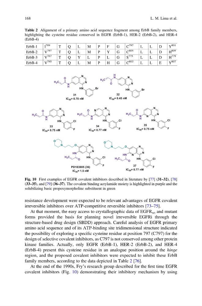
At the end of the 1990s, Fry’s research group described for the first time EGFR
covalent inhibitors (Fig. 10) demonstrating their inhibitory mechanism by using
Table 2 Alignment of a primary amino acid sequence fragment among ErbB family members,
highlighting the cysteine residue conserved in EGFR (ErbB-1), HER-2 (ErbB-2), and HER-4
(ErbB-4)
ErbB-1
I
789
T
Q
L
M
P
F
G
C
797
L
L
D
Y
801
ErbB-2
V
797
T
Q
L
M
P
Y
G
C
805
L
L
D
H
809
ErbB-3
V
767
T
Q
Y
L
P
L
G
S
775
L
L
D
H
779
ErbB-4
V
795
T
Q
L
M
P
H
G
C
803
L
L
E
Y
807
Fig. 10 First examples of EGFR covalent inhibitors described in literature by [77] (31–32), [78]
(33–35), and [79] (36–37). The covalent binding acrylamide moiety is highlighted in purple and the
solubilizing basic propoxymorpholine substituent in green
168
L. M. Lima et al.
irreversible inhibitors over ATP-competitive reversible inhibitors [73–75].
At that moment, the easy access to crystallographic data of EGFR wt and mutant
forms provided the basis for planning novel irreversible EGFRi through the
structure-based drug design (SBDD) approach. Careful analysis of EGFR primary
amino acid sequence and of its ATP-binding site tridimensional structure indicated
the possibility of exploring a specific cysteine residue at position 797 (C797) for the
design of selective covalent inhibitors, as C797 is not conserved among other protein
kinase families. Actually, only EGFR (ErbB-1), HER-2 (ErbB-2), and HER-4
(ErbB-4) present this cysteine residue in an analogue position around the hinge
region, and the proposed covalent inhibitors were expected to inhibit these ErbB
family members, according to the data depicted in Table 2 [76].
At the end of the 1990s, Fry’s research group described for the first time EGFR
covalent inhibitors (Fig. 10) demonstrating their inhibitory mechanism by using
Table 2 Alignment of a primary amino acid sequence fragment among ErbB family members,
highlighting the cysteine residue conserved in EGFR (ErbB-1), HER-2 (ErbB-2), and HER-4
(ErbB-4)
ErbB-1
I
789
T
Q
L
M
P
F
G
C
797
L
L
D
Y
801
ErbB-2
V
797
T
Q
L
M
P
Y
G
C
805
L
L
D
H
809
ErbB-3
V
767
T
Q
Y
L
P
L
G
S
775
L
L
D
H
779
ErbB-4
V
795
T
Q
L
M
P
H
G
C
803
L
L
E
Y
807
Fig. 10 First examples of EGFR covalent inhibitors described in literature by [77] (31–32), [78]
(33–35), and [79] (36–37). The covalent binding acrylamide moiety is highlighted in purple and the
solubilizing basic propoxymorpholine substituent in green
168
L. M. Lima et al.