

Because of the structural diversity that is also often observed in active
kinases, Taylor et al. established a better definition of the active state by analysis
of hydrophobic spines that bridge both kinase lobes and interconnect all elements
important for activity. The spines are present in all active kinases, and this structural
feature seems therefore better suited defining the active state. Consequently,
in inactive kinases the spines are broken leaving one of more structural motif
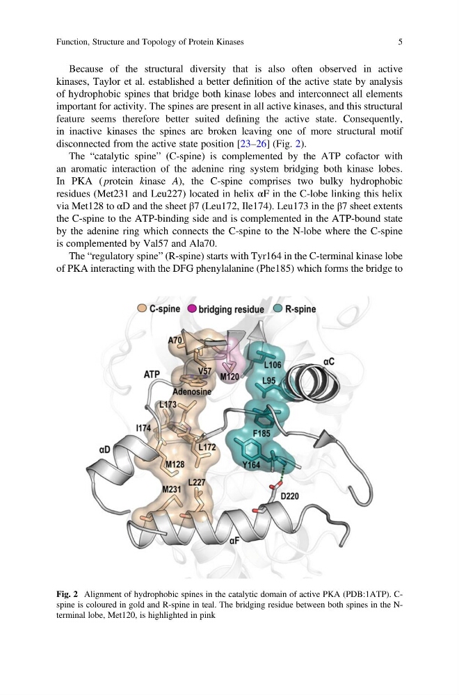
disconnected from the active state position [23–26] (Fig. 2).
The “catalytic spine” (C-spine) is complemented by the ATP cofactor with
an aromatic interaction of the adenine ring system bridging both kinase lobes.
In PKA ( protein kinase A), the C-spine comprises two bulky hydrophobic
residues (Met231 and Leu227) located in helix αF in the C-lobe linking this helix
via Met128 to αD and the sheet β7 (Leu172, Ile174). Leu173 in the β7 sheet extents
the C-spine to the ATP-binding side and is complemented in the ATP-bound state
by the adenine ring which connects the C-spine to the N-lobe where the C-spine
is complemented by Val57 and Ala70.
The “regulatory spine” (R-spine) starts with Tyr164 in the C-terminal kinase lobe
of PKA interacting with the DFG phenylalanine (Phe185) which forms the bridge to
Fig. 2 Alignment of hydrophobic spines in the catalytic domain of active PKA (PDB:1ATP). Cspine is coloured in gold and R-spine in teal. The bridging residue between both spines in the Nterminal lobe, Met120, is highlighted in pink
Function, Structure and Topology of Protein Kinases
5
kinases, Taylor et al. established a better definition of the active state by analysis
of hydrophobic spines that bridge both kinase lobes and interconnect all elements
important for activity. The spines are present in all active kinases, and this structural
feature seems therefore better suited defining the active state. Consequently,
in inactive kinases the spines are broken leaving one of more structural motif
disconnected from the active state position [23–26] (Fig. 2).
The “catalytic spine” (C-spine) is complemented by the ATP cofactor with
an aromatic interaction of the adenine ring system bridging both kinase lobes.
In PKA ( protein kinase A), the C-spine comprises two bulky hydrophobic
residues (Met231 and Leu227) located in helix αF in the C-lobe linking this helix
via Met128 to αD and the sheet β7 (Leu172, Ile174). Leu173 in the β7 sheet extents
the C-spine to the ATP-binding side and is complemented in the ATP-bound state
by the adenine ring which connects the C-spine to the N-lobe where the C-spine
is complemented by Val57 and Ala70.
The “regulatory spine” (R-spine) starts with Tyr164 in the C-terminal kinase lobe
of PKA interacting with the DFG phenylalanine (Phe185) which forms the bridge to
Fig. 2 Alignment of hydrophobic spines in the catalytic domain of active PKA (PDB:1ATP). Cspine is coloured in gold and R-spine in teal. The bridging residue between both spines in the Nterminal lobe, Met120, is highlighted in pink
Function, Structure and Topology of Protein Kinases
5