

4.4 Zinc-Based Nanomaterials and Hydrogen Sulfide Decomposition
43
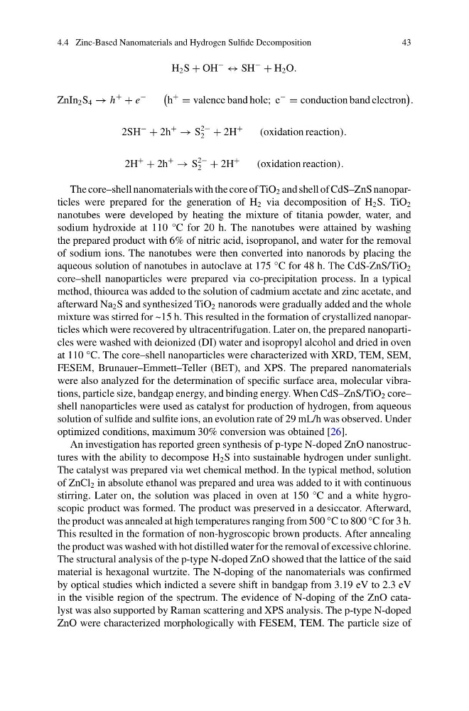
H 2 S + OH
−
↔ SH
−
+ H 2 O.
ZnIn 2 S 4 → h
+
+ e
−
h
+
= valence band hole; e
−
= conduction band electron
.
2SH
−
+ 2h
+
→ S
2−
2 + 2H
+
(oxidation reaction).
2H
+
+ 2h
+
→ S
2−
2 + 2H
+
(oxidation reaction).
The core–shell nanomaterials with the core of TiO 2 and shell of CdS–ZnS nanoparticles were prepared for the generation of H 2 via decomposition of H 2 S. TiO 2
nanotubes were developed by heating the mixture of titania powder, water, and
sodium hydroxide at 110 °C for 20 h. The nanotubes were attained by washing
the prepared product with 6% of nitric acid, isopropanol, and water for the removal
of sodium ions. The nanotubes were then converted into nanorods by placing the
aqueous solution of nanotubes in autoclave at 175 °C for 48 h. The CdS-ZnS/TiO 2
core–shell nanoparticles were prepared via co-precipitation process. In a typical
method, thiourea was added to the solution of cadmium acetate and zinc acetate, and
afterward Na 2 S and synthesized TiO 2 nanorods were gradually added and the whole
mixture was stirred for ~15 h. This resulted in the formation of crystallized nanoparticles which were recovered by ultracentrifugation. Later on, the prepared nanoparticles were washed with deionized (DI) water and isopropyl alcohol and dried in oven
at 110 °C. The core–shell nanoparticles were characterized with XRD, TEM, SEM,
FESEM, Brunauer–Emmett–Teller (BET), and XPS. The prepared nanomaterials
were also analyzed for the determination of specific surface area, molecular vibrations, particle size, bandgap energy, and binding energy. When CdS–ZnS/TiO 2 core–
shell nanoparticles were used as catalyst for production of hydrogen, from aqueous
solution of sulfide and sulfite ions, an evolution rate of 29 mL/h was observed. Under
optimized conditions, maximum 30% conversion was obtained [26].
An investigation has reported green synthesis of p-type N-doped ZnO nanostructures with the ability to decompose H 2 S into sustainable hydrogen under sunlight.
The catalyst was prepared via wet chemical method. In the typical method, solution
of ZnCl 2 in absolute ethanol was prepared and urea was added to it with continuous
stirring. Later on, the solution was placed in oven at 150 °C and a white hygroscopic product was formed. The product was preserved in a desiccator. Afterward,
the product was annealed at high temperatures ranging from 500 °C to 800 °C for 3 h.
This resulted in the formation of non-hygroscopic brown products. After annealing
the product was washed with hot distilled water for the removal of excessive chlorine.
The structural analysis of the p-type N-doped ZnO showed that the lattice of the said
material is hexagonal wurtzite. The N-doping of the nanomaterials was confirmed
by optical studies which indicted a severe shift in bandgap from 3.19 eV to 2.3 eV
in the visible region of the spectrum. The evidence of N-doping of the ZnO catalyst was also supported by Raman scattering and XPS analysis. The p-type N-doped
ZnO were characterized morphologically with FESEM, TEM. The particle size of
43
H 2 S + OH
−
↔ SH
−
+ H 2 O.
ZnIn 2 S 4 → h
+
+ e
−
h
+
= valence band hole; e
−
= conduction band electron
.
2SH
−
+ 2h
+
→ S
2−
2 + 2H
+
(oxidation reaction).
2H
+
+ 2h
+
→ S
2−
2 + 2H
+
(oxidation reaction).
The core–shell nanomaterials with the core of TiO 2 and shell of CdS–ZnS nanoparticles were prepared for the generation of H 2 via decomposition of H 2 S. TiO 2
nanotubes were developed by heating the mixture of titania powder, water, and
sodium hydroxide at 110 °C for 20 h. The nanotubes were attained by washing
the prepared product with 6% of nitric acid, isopropanol, and water for the removal
of sodium ions. The nanotubes were then converted into nanorods by placing the
aqueous solution of nanotubes in autoclave at 175 °C for 48 h. The CdS-ZnS/TiO 2
core–shell nanoparticles were prepared via co-precipitation process. In a typical
method, thiourea was added to the solution of cadmium acetate and zinc acetate, and
afterward Na 2 S and synthesized TiO 2 nanorods were gradually added and the whole
mixture was stirred for ~15 h. This resulted in the formation of crystallized nanoparticles which were recovered by ultracentrifugation. Later on, the prepared nanoparticles were washed with deionized (DI) water and isopropyl alcohol and dried in oven
at 110 °C. The core–shell nanoparticles were characterized with XRD, TEM, SEM,
FESEM, Brunauer–Emmett–Teller (BET), and XPS. The prepared nanomaterials
were also analyzed for the determination of specific surface area, molecular vibrations, particle size, bandgap energy, and binding energy. When CdS–ZnS/TiO 2 core–
shell nanoparticles were used as catalyst for production of hydrogen, from aqueous
solution of sulfide and sulfite ions, an evolution rate of 29 mL/h was observed. Under
optimized conditions, maximum 30% conversion was obtained [26].
An investigation has reported green synthesis of p-type N-doped ZnO nanostructures with the ability to decompose H 2 S into sustainable hydrogen under sunlight.
The catalyst was prepared via wet chemical method. In the typical method, solution
of ZnCl 2 in absolute ethanol was prepared and urea was added to it with continuous
stirring. Later on, the solution was placed in oven at 150 °C and a white hygroscopic product was formed. The product was preserved in a desiccator. Afterward,
the product was annealed at high temperatures ranging from 500 °C to 800 °C for 3 h.
This resulted in the formation of non-hygroscopic brown products. After annealing
the product was washed with hot distilled water for the removal of excessive chlorine.
The structural analysis of the p-type N-doped ZnO showed that the lattice of the said
material is hexagonal wurtzite. The N-doping of the nanomaterials was confirmed
by optical studies which indicted a severe shift in bandgap from 3.19 eV to 2.3 eV
in the visible region of the spectrum. The evidence of N-doping of the ZnO catalyst was also supported by Raman scattering and XPS analysis. The p-type N-doped
ZnO were characterized morphologically with FESEM, TEM. The particle size of