

62
A. Jayaraman et al.
the matrix. [2] This is followed by a description of the simulation protocol used to
study a single polymer grafted particle in a polymer matrix and the relevant structural
analyses. We present some key results for a select few systems and conclude with a
discussion of future directions.
4.2 Model
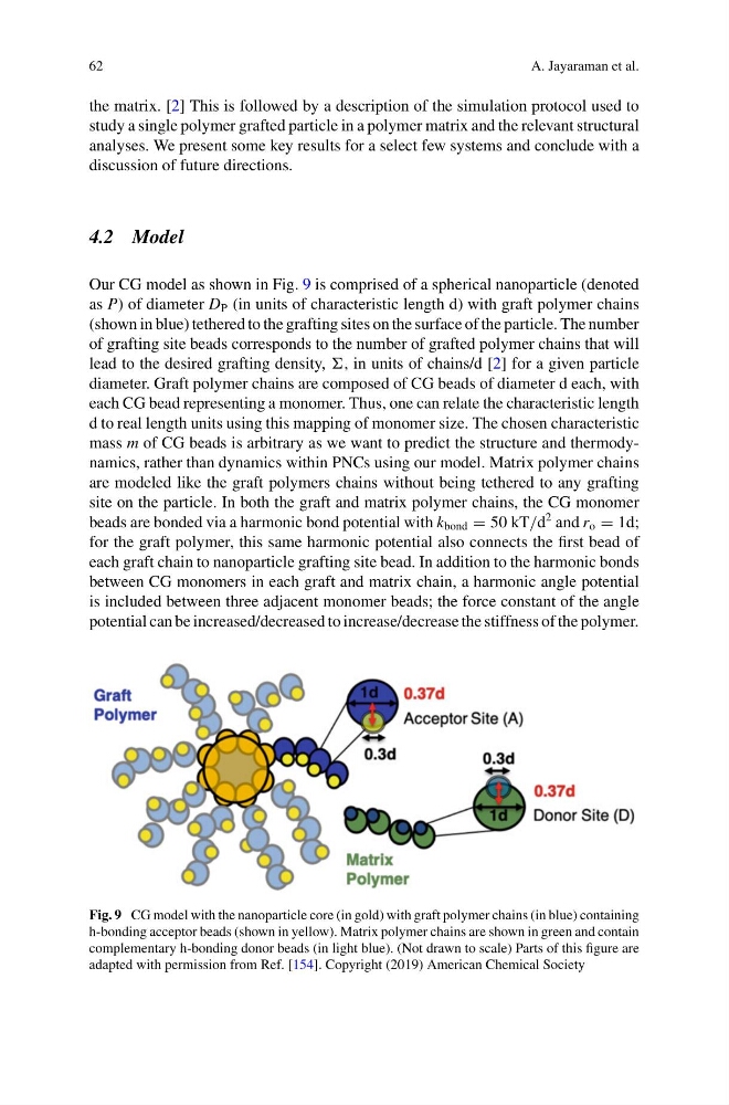
Our CG model as shown in Fig. 9 is comprised of a spherical nanoparticle (denoted
as P) of diameter D P (in units of characteristic length d) with graft polymer chains
(shown in blue) tethered to the grafting sites on the surface of the particle. The number
of grafting site beads corresponds to the number of grafted polymer chains that will
lead to the desired grafting density, , in units of chains/d [2] for a given particle
diameter. Graft polymer chains are composed of CG beads of diameter d each, with
each CG bead representing a monomer. Thus, one can relate the characteristic length
d to real length units using this mapping of monomer size. The chosen characteristic
mass m of CG beads is arbitrary as we want to predict the structure and thermodynamics, rather than dynamics within PNCs using our model. Matrix polymer chains
are modeled like the graft polymers chains without being tethered to any grafting
site on the particle. In both the graft and matrix polymer chains, the CG monomer
beads are bonded via a harmonic bond potential with k bond = 50 kT/d
2 and r o = 1d;
for the graft polymer, this same harmonic potential also connects the first bead of
each graft chain to nanoparticle grafting site bead. In addition to the harmonic bonds
between CG monomers in each graft and matrix chain, a harmonic angle potential
is included between three adjacent monomer beads; the force constant of the angle
potential can be increased/decreased to increase/decrease the stiffness of the polymer.
Fig. 9 CG model with the nanoparticle core (in gold) with graft polymer chains (in blue) containing
h-bonding acceptor beads (shown in yellow). Matrix polymer chains are shown in green and contain
complementary h-bonding donor beads (in light blue). (Not drawn to scale) Parts of this figure are
adapted with permission from Ref. [154]. Copyright (2019) American Chemical Society
A. Jayaraman et al.
the matrix. [2] This is followed by a description of the simulation protocol used to
study a single polymer grafted particle in a polymer matrix and the relevant structural
analyses. We present some key results for a select few systems and conclude with a
discussion of future directions.
4.2 Model
Our CG model as shown in Fig. 9 is comprised of a spherical nanoparticle (denoted
as P) of diameter D P (in units of characteristic length d) with graft polymer chains
(shown in blue) tethered to the grafting sites on the surface of the particle. The number
of grafting site beads corresponds to the number of grafted polymer chains that will
lead to the desired grafting density, , in units of chains/d [2] for a given particle
diameter. Graft polymer chains are composed of CG beads of diameter d each, with
each CG bead representing a monomer. Thus, one can relate the characteristic length
d to real length units using this mapping of monomer size. The chosen characteristic
mass m of CG beads is arbitrary as we want to predict the structure and thermodynamics, rather than dynamics within PNCs using our model. Matrix polymer chains
are modeled like the graft polymers chains without being tethered to any grafting
site on the particle. In both the graft and matrix polymer chains, the CG monomer
beads are bonded via a harmonic bond potential with k bond = 50 kT/d
2 and r o = 1d;
for the graft polymer, this same harmonic potential also connects the first bead of
each graft chain to nanoparticle grafting site bead. In addition to the harmonic bonds
between CG monomers in each graft and matrix chain, a harmonic angle potential
is included between three adjacent monomer beads; the force constant of the angle
potential can be increased/decreased to increase/decrease the stiffness of the polymer.
Fig. 9 CG model with the nanoparticle core (in gold) with graft polymer chains (in blue) containing
h-bonding acceptor beads (shown in yellow). Matrix polymer chains are shown in green and contain
complementary h-bonding donor beads (in light blue). (Not drawn to scale) Parts of this figure are
adapted with permission from Ref. [154]. Copyright (2019) American Chemical Society