

34
S. Takaichi
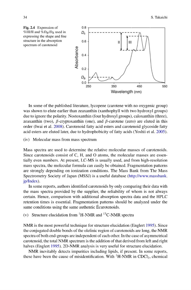
Fig. 2.4 Expression of
%III/II and %D B /D II used in
expressing the shape and fine
structure in the absorption
spectrum of carotenoid
In some of the published literature, lycopene (carotene with no oxygenic group)
was shown to elute earlier than zeaxanthin (xanthophyll with two hydroxyl groups)
due to ignore the polarity. Nostoxanthin (four hydroxyl groups), caloxanthin (three),
zeaxanthin (two), β-cryptoxanthin (one), and β-carotene (zero) are eluted in this
order (Iwai et al. 2008). Carotenoid fatty acid esters and carotenoid glycoside fatty
acid esters are eluted later, due to hydrophobicity of fatty acids (Yoshii et al. 2005).
(iv) Molecular mass from mass spectrum
Mass spectra are used to determine the relative molecular masses of carotenoids.
Since carotenoids consist of C, H, and O atoms, the molecular masses are essentially even numbers. At present, LC-MS is usually used, and from high-resolution
mass spectra, the molecular formula can easily be obtained. Fragmentation patterns
are strongly depending on ionization conditions. The Mass Bank from The Mass
Spectrometry Society of Japan (MSSJ) is a useful database (http://www.massbank.
jp/Index).
In some reports, authors identified carotenoids by only comparing their data with
the mass spectra provided by the supplier, the reliability of whom is not always
certain. Hence, comparison with additional absorption spectra data and the HPLC
retention times is essential. Fragmentation patterns should be analyzed under the
same conditions using the same authentic Écarotenoids.
(v) Structure elucidation from
1 H-NMR and
13 C-NMR spectra
NMR is the most powerful technique for structure elucidation (Englert 1995). Since
the conjugated double bonds of the olefinic region of carotenoids are long, the NMR
spectra of both end-groups are independent of each other. In the case of asymmetrical
carotenoid, the total NMR spectrum is the addition of that derived from left and right
halves (Englert 1995). 2D-NMR analysis is very useful for structure elucidation.
NMR inevitably detects impurities including lipids, if present. In some reports,
these have been the cause of misidentification. With
1 H-NMR in CDCl 3 , chemical
S. Takaichi
Fig. 2.4 Expression of
%III/II and %D B /D II used in
expressing the shape and fine
structure in the absorption
spectrum of carotenoid
In some of the published literature, lycopene (carotene with no oxygenic group)
was shown to elute earlier than zeaxanthin (xanthophyll with two hydroxyl groups)
due to ignore the polarity. Nostoxanthin (four hydroxyl groups), caloxanthin (three),
zeaxanthin (two), β-cryptoxanthin (one), and β-carotene (zero) are eluted in this
order (Iwai et al. 2008). Carotenoid fatty acid esters and carotenoid glycoside fatty
acid esters are eluted later, due to hydrophobicity of fatty acids (Yoshii et al. 2005).
(iv) Molecular mass from mass spectrum
Mass spectra are used to determine the relative molecular masses of carotenoids.
Since carotenoids consist of C, H, and O atoms, the molecular masses are essentially even numbers. At present, LC-MS is usually used, and from high-resolution
mass spectra, the molecular formula can easily be obtained. Fragmentation patterns
are strongly depending on ionization conditions. The Mass Bank from The Mass
Spectrometry Society of Japan (MSSJ) is a useful database (http://www.massbank.
jp/Index).
In some reports, authors identified carotenoids by only comparing their data with
the mass spectra provided by the supplier, the reliability of whom is not always
certain. Hence, comparison with additional absorption spectra data and the HPLC
retention times is essential. Fragmentation patterns should be analyzed under the
same conditions using the same authentic Écarotenoids.
(v) Structure elucidation from
1 H-NMR and
13 C-NMR spectra
NMR is the most powerful technique for structure elucidation (Englert 1995). Since
the conjugated double bonds of the olefinic region of carotenoids are long, the NMR
spectra of both end-groups are independent of each other. In the case of asymmetrical
carotenoid, the total NMR spectrum is the addition of that derived from left and right
halves (Englert 1995). 2D-NMR analysis is very useful for structure elucidation.
NMR inevitably detects impurities including lipids, if present. In some reports,
these have been the cause of misidentification. With
1 H-NMR in CDCl 3 , chemical