

5 Carotenoid Overproduction in Microalgae: Biochemical …
107
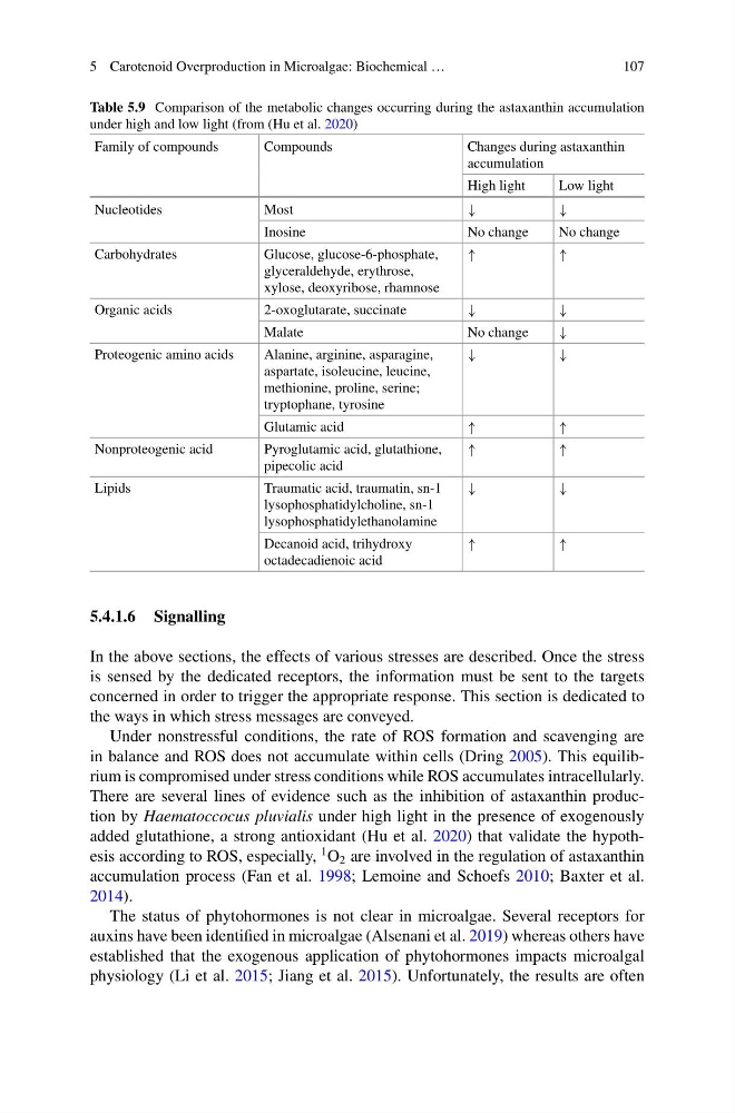
Table 5.9 Comparison of the metabolic changes occurring during the astaxanthin accumulation
under high and low light (from (Hu et al. 2020)
Family of compounds
Compounds
Changes during astaxanthin
accumulation
High light
Low light
Nucleotides
Most
↓
↓
Inosine
No change
No change
Carbohydrates
Glucose, glucose-6-phosphate,
glyceraldehyde, erythrose,
xylose, deoxyribose, rhamnose
↑
↑
Organic acids
2-oxoglutarate, succinate
↓
↓
Malate
No change
↓
Proteogenic amino acids
Alanine, arginine, asparagine,
aspartate, isoleucine, leucine,
methionine, proline, serine;
tryptophane, tyrosine
↓
↓
Glutamic acid
↑
↑
Nonproteogenic acid
Pyroglutamic acid, glutathione,
pipecolic acid
↑
↑
Lipids
Traumatic acid, traumatin, sn-1
lysophosphatidylcholine, sn-1
lysophosphatidylethanolamine
↓
↓
Decanoid acid, trihydroxy
octadecadienoic acid
↑
↑
5.4.1.6 Signalling
In the above sections, the effects of various stresses are described. Once the stress
is sensed by the dedicated receptors, the information must be sent to the targets
concerned in order to trigger the appropriate response. This section is dedicated to
the ways in which stress messages are conveyed.
Under nonstressful conditions, the rate of ROS formation and scavenging are
in balance and ROS does not accumulate within cells (Dring 2005). This equilibrium is compromised under stress conditions while ROS accumulates intracellularly.
There are several lines of evidence such as the inhibition of astaxanthin production by Haematoccocus pluvialis under high light in the presence of exogenously
added glutathione, a strong antioxidant (Hu et al. 2020) that validate the hypothesis according to ROS, especially,
1 O 2 are involved in the regulation of astaxanthin
accumulation process (Fan et al. 1998; Lemoine and Schoefs 2010; Baxter et al.
2014).
The status of phytohormones is not clear in microalgae. Several receptors for
auxins have been identified in microalgae (Alsenani et al. 2019) whereas others have
established that the exogenous application of phytohormones impacts microalgal
physiology (Li et al. 2015; Jiang et al. 2015). Unfortunately, the results are often
107
Table 5.9 Comparison of the metabolic changes occurring during the astaxanthin accumulation
under high and low light (from (Hu et al. 2020)
Family of compounds
Compounds
Changes during astaxanthin
accumulation
High light
Low light
Nucleotides
Most
↓
↓
Inosine
No change
No change
Carbohydrates
Glucose, glucose-6-phosphate,
glyceraldehyde, erythrose,
xylose, deoxyribose, rhamnose
↑
↑
Organic acids
2-oxoglutarate, succinate
↓
↓
Malate
No change
↓
Proteogenic amino acids
Alanine, arginine, asparagine,
aspartate, isoleucine, leucine,
methionine, proline, serine;
tryptophane, tyrosine
↓
↓
Glutamic acid
↑
↑
Nonproteogenic acid
Pyroglutamic acid, glutathione,
pipecolic acid
↑
↑
Lipids
Traumatic acid, traumatin, sn-1
lysophosphatidylcholine, sn-1
lysophosphatidylethanolamine
↓
↓
Decanoid acid, trihydroxy
octadecadienoic acid
↑
↑
5.4.1.6 Signalling
In the above sections, the effects of various stresses are described. Once the stress
is sensed by the dedicated receptors, the information must be sent to the targets
concerned in order to trigger the appropriate response. This section is dedicated to
the ways in which stress messages are conveyed.
Under nonstressful conditions, the rate of ROS formation and scavenging are
in balance and ROS does not accumulate within cells (Dring 2005). This equilibrium is compromised under stress conditions while ROS accumulates intracellularly.
There are several lines of evidence such as the inhibition of astaxanthin production by Haematoccocus pluvialis under high light in the presence of exogenously
added glutathione, a strong antioxidant (Hu et al. 2020) that validate the hypothesis according to ROS, especially,
1 O 2 are involved in the regulation of astaxanthin
accumulation process (Fan et al. 1998; Lemoine and Schoefs 2010; Baxter et al.
2014).
The status of phytohormones is not clear in microalgae. Several receptors for
auxins have been identified in microalgae (Alsenani et al. 2019) whereas others have
established that the exogenous application of phytohormones impacts microalgal
physiology (Li et al. 2015; Jiang et al. 2015). Unfortunately, the results are often