

wide diversity of adult organs, most of which comprise a mixture of
epithelial cells and supporting mesenchymal cells in close association. Thus, throughout development, epithelial cells arise from
non-epithelial cell types through MET.
Mesodermal cells along the mediolateral axis undergo MET to
generate the epithelial precursors of the notochord, somites,
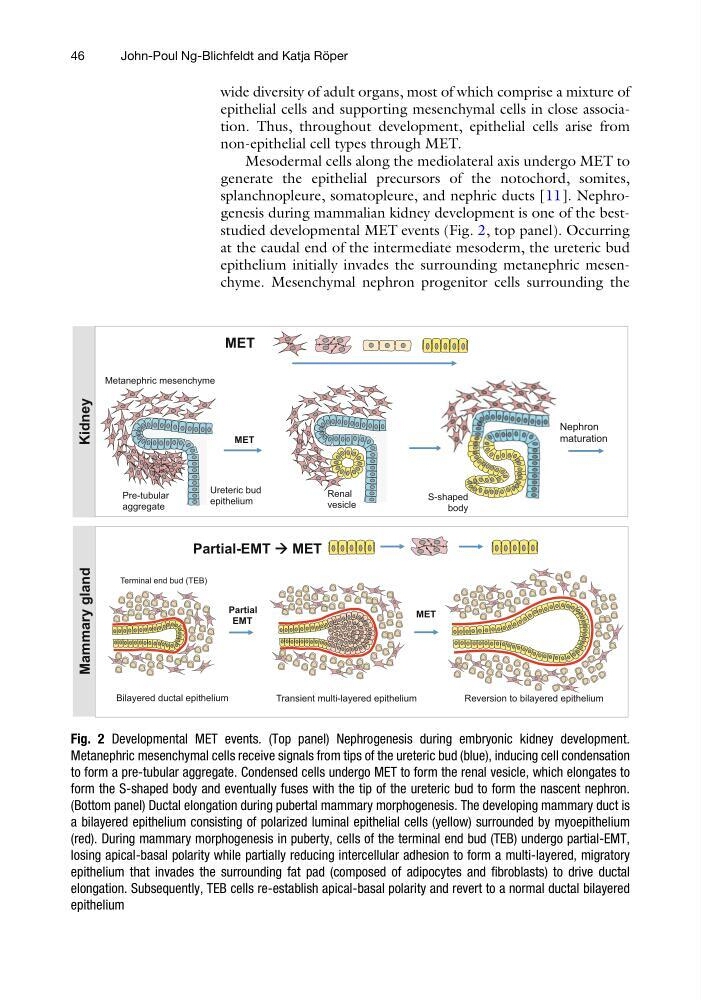
splanchnopleure, somatopleure, and nephric ducts [11]. Nephrogenesis during mammalian kidney development is one of the beststudied developmental MET events (Fig. 2, top panel). Occurring
at the caudal end of the intermediate mesoderm, the ureteric bud
epithelium initially invades the surrounding metanephric mesenchyme. Mesenchymal nephron progenitor cells surrounding the
Mammary gland
Renal
vesicle
Metanephric mesenchyme
Pre-tubular
aggregate
Ureteric bud
epithelium
Kidney
MET
S-shaped
body
Nephron
maturation
` ` ` ` `
`
`
`
`
`
`
`
`
`
`
MET
Partial-EMT
MET
MET
Partial
EMT
Transient multi-layered epithelium
Reversion to bilayered epithelium
Bilayered ductal epithelium
Terminal end bud (TEB)
Fig. 2 Developmental MET events. (Top panel) Nephrogenesis during embryonic kidney development.
Metanephric mesenchymal cells receive signals from tips of the ureteric bud (blue), inducing cell condensation
to form a pre-tubular aggregate. Condensed cells undergo MET to form the renal vesicle, which elongates to
form the S-shaped body and eventually fuses with the tip of the ureteric bud to form the nascent nephron.
(Bottom panel) Ductal elongation during pubertal mammary morphogenesis. The developing mammary duct is
a bilayered epithelium consisting of polarized luminal epithelial cells (yellow) surrounded by myoepithelium
(red). During mammary morphogenesis in puberty, cells of the terminal end bud (TEB) undergo partial-EMT,
losing apical-basal polarity while partially reducing intercellular adhesion to form a multi-layered, migratory
epithelium that invades the surrounding fat pad (composed of adipocytes and fibroblasts) to drive ductal
elongation. Subsequently, TEB cells re-establish apical-basal polarity and revert to a normal ductal bilayered
epithelium
46
John-Poul Ng-Blichfeldt and Katja Ro ¨ per
epithelial cells and supporting mesenchymal cells in close association. Thus, throughout development, epithelial cells arise from
non-epithelial cell types through MET.
Mesodermal cells along the mediolateral axis undergo MET to
generate the epithelial precursors of the notochord, somites,
splanchnopleure, somatopleure, and nephric ducts [11]. Nephrogenesis during mammalian kidney development is one of the beststudied developmental MET events (Fig. 2, top panel). Occurring
at the caudal end of the intermediate mesoderm, the ureteric bud
epithelium initially invades the surrounding metanephric mesenchyme. Mesenchymal nephron progenitor cells surrounding the
Mammary gland
Renal
vesicle
Metanephric mesenchyme
Pre-tubular
aggregate
Ureteric bud
epithelium
Kidney
MET
S-shaped
body
Nephron
maturation
` ` ` ` `
`
`
`
`
`
`
`
`
`
`
MET
Partial-EMT
MET
MET
Partial
EMT
Transient multi-layered epithelium
Reversion to bilayered epithelium
Bilayered ductal epithelium
Terminal end bud (TEB)
Fig. 2 Developmental MET events. (Top panel) Nephrogenesis during embryonic kidney development.
Metanephric mesenchymal cells receive signals from tips of the ureteric bud (blue), inducing cell condensation
to form a pre-tubular aggregate. Condensed cells undergo MET to form the renal vesicle, which elongates to
form the S-shaped body and eventually fuses with the tip of the ureteric bud to form the nascent nephron.
(Bottom panel) Ductal elongation during pubertal mammary morphogenesis. The developing mammary duct is
a bilayered epithelium consisting of polarized luminal epithelial cells (yellow) surrounded by myoepithelium
(red). During mammary morphogenesis in puberty, cells of the terminal end bud (TEB) undergo partial-EMT,
losing apical-basal polarity while partially reducing intercellular adhesion to form a multi-layered, migratory
epithelium that invades the surrounding fat pad (composed of adipocytes and fibroblasts) to drive ductal
elongation. Subsequently, TEB cells re-establish apical-basal polarity and revert to a normal ductal bilayered
epithelium
46
John-Poul Ng-Blichfeldt and Katja Ro ¨ per