

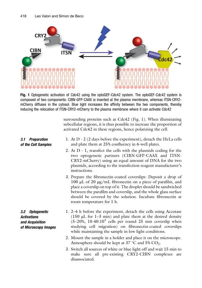
surrounding proteins such as Cdc42 (Fig. 1). When illuminating
subcellular regions, it is thus possible to increase the proportion of
activated Cdc42 in these regions, hence polarizing the cell.
3.1 Preparation
of the Cell Samples
1. At D - 2 (2 days before the experiment), detach the HeLa cells
and plate them at 25% confluency in 6-well plates.
2. At D - 1, transfect the cells with the plasmids coding for the
two optogenetic partners (CIBN-GFP-CAAX and ITSNCRY2-mCherry) using an equal amount of DNA for the two
plasmids, according to the transfection reagent manufacturer’s
instructions.
3. Prepare the fibronectin-coated coverslips: Deposit a drop of
100 μL of 20 μg/mL fibronectin on a piece of parafilm, and
place a coverslip on top of it. The droplet should be sandwiched
between the parafilm and coverslip, and the whole glass surface
should be covered by the solution. Incubate fibronectin at
room temperature for 1 h.
3.2 Optogenetic
Activations
and Acquisition
of Microscopy Images
1. 2–4 h before the experiment, detach the cells using Accutase
(150 μL for 1–3 min) and plate them at the desired density
(5–20%, 10-40.10
3 cells per round 25 mm coverslip when
studying cell migration) on fibronectin-coated coverslips
while maintaining the sample in low light conditions.
2. Mount the sample in a holder and place it on the microscope.
Atmosphere should be kept at 37
C and 5% CO 2 .
3. Switch all sources of white or blue light off and wait 15 min to
make sure all pre-existing CRY2-CIBN complexes are
disassociated.
Fig. 1 Optogenetic activation of Cdc42 using the optoGEF-Cdc42 system. The optoGEF-Cdc42 system is
composed of two components: CIBN-GFP-CAAX is inserted at the plasma membrane, whereas ITSN-CRY2mCherry diffuses in the cytosol. Blue light increases the affinity between the two components, thereby
inducing the relocation of ITSN-CRY2-mCherry to the plasma membrane where it can activate Cdc42
418
Leo Valon and Simon de Beco
subcellular regions, it is thus possible to increase the proportion of
activated Cdc42 in these regions, hence polarizing the cell.
3.1 Preparation
of the Cell Samples
1. At D - 2 (2 days before the experiment), detach the HeLa cells
and plate them at 25% confluency in 6-well plates.
2. At D - 1, transfect the cells with the plasmids coding for the
two optogenetic partners (CIBN-GFP-CAAX and ITSNCRY2-mCherry) using an equal amount of DNA for the two
plasmids, according to the transfection reagent manufacturer’s
instructions.
3. Prepare the fibronectin-coated coverslips: Deposit a drop of
100 μL of 20 μg/mL fibronectin on a piece of parafilm, and
place a coverslip on top of it. The droplet should be sandwiched
between the parafilm and coverslip, and the whole glass surface
should be covered by the solution. Incubate fibronectin at
room temperature for 1 h.
3.2 Optogenetic
Activations
and Acquisition
of Microscopy Images
1. 2–4 h before the experiment, detach the cells using Accutase
(150 μL for 1–3 min) and plate them at the desired density
(5–20%, 10-40.10
3 cells per round 25 mm coverslip when
studying cell migration) on fibronectin-coated coverslips
while maintaining the sample in low light conditions.
2. Mount the sample in a holder and place it on the microscope.
Atmosphere should be kept at 37
C and 5% CO 2 .
3. Switch all sources of white or blue light off and wait 15 min to
make sure all pre-existing CRY2-CIBN complexes are
disassociated.
Fig. 1 Optogenetic activation of Cdc42 using the optoGEF-Cdc42 system. The optoGEF-Cdc42 system is
composed of two components: CIBN-GFP-CAAX is inserted at the plasma membrane, whereas ITSN-CRY2mCherry diffuses in the cytosol. Blue light increases the affinity between the two components, thereby
inducing the relocation of ITSN-CRY2-mCherry to the plasma membrane where it can activate Cdc42
418
Leo Valon and Simon de Beco